Download
1 / 4
40 likes | 114 Views
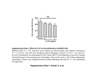
This study investigates the effects of LL-37 peptide on HMVEC-LBls proliferation and LPS receptor expression. Results show LL-37 modulation of cell proliferation and LPS receptor levels. The data suggest potential roles of LL-37 in immune response regulation.

E N D
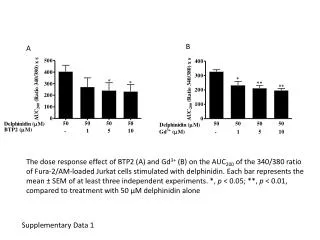
LL-37 (mg/ml) Supplementary Data 1. Effect of LL-37 on the proliferation of HMVEC-LBls. HMVEC-LBls (4 × 10⁴ cells/cm2) were seeded into 48-well plates and cultured overnight at 37°C, 5% CO2. The cells were incubated without (Resting) or with LL-37 (0.1, 1 or 5 mg ml-1) for 24 h. Proliferation was determined by MTT assay (MTT Cell Proliferation Assay Kit, Cayman Chemical) based on the manufacturer’s description. Data are the mean SE of three independent experiments. Values were compared between without (Resting) and with LL-37. NS; statistically not significant. Supplementary Data 1. Suzuki, K. et al.
CD14 Resting LL-37 LPS/CHX LPS/CHX+LL-37 Isotype IgG 500 500 500 500 500 500 500 500 TLR4 aCD14 mAb 400 400 400 400 400 400 400 400 300 300 300 300 300 300 300 300 Cell counts Cell counts Cell counts Cell counts Cell counts Cell counts Cell counts Cell counts 200 200 200 200 200 200 200 200 100 100 100 100 100 100 100 100 0 0 0 0 0 0 0 0 FL-2 FL-2 FL-2 FL-2 FL-2 FL-2 FL-2 FL-2 Isotype IgG aTLR4 mAb Supplementary Data 2. Effects of LL-37 on the expression of LPS receptors on HMVEC-LBls. HMVEC-LBls were seeded into 6-well plates and cultured overnight at 37°C, 5% CO2. Semi-confluent HMVEC-LBls were incubated without (Resting), with LL-37 (1 mg ml-1) alone, or with LPS (100 ng ml-1) and CHX (10 mg ml-1) (LPS/CHX) in the absence or presence of LL-37 (+LL-37; 1 mg ml-1) for 24 h. Cells were detached with 0.05% trypsin, 0.01% EDTA/PBS, suspended in 0.5% BSA/PBS and incubated with phycoerythrin-conjugated anti-human CD14 mAb (MY-4), anti-human TLR4 mAb (HTA125) or isotype IgG (2.5 mg ml-1) for 30 min on ice. Cells were washed, resuspended in 0.5% BSA/PBS, and then analyzed by flow cytometry. Data are representative of two independent experiments. Supplementary Data 2. Suzuki, K. et al.
CD14 TLR4 Isotype IgG Isotype IgG 500 500 aTLR4 mAb aCD14 mAb 400 400 300 300 Cell counts Cell counts 200 200 100 100 0 0 Supplementary Data 3. Expression of LPS receptors on mouse hepatic endothelial cells. Mouse liver was perfused in situ via the portal vein with 0.05% collagenase type IV (Sigma-Aldrich) according to the previous report (43). Hepatic non-parenchymal cells containing endothelial cells, Kupffer cells, stellate cells and blood cells were separated from hepatocytes by low-speed centrifugation. The hemolysed non-parenchymal cells were blocked with anti-mouse Fcg Receptor mAb (2.4G2, Becton Dickinson), and stained with biotinylated anti-mouse CD31 mAb (MEC 13.3) and phycoerythrin-conjugated anti-mouse CD14 mAb (Sa2-8, eBioscience) or anti-mouse TLR4 mAb (UT41, eBioscience) (2.5 mg ml-1). Cells were further stained with allophycocyanin-conjugated streptavidin, washed, resuspended in 0.5% BSA/PBS, and then analyzed by flow cytometry (FACSCalibur, Becton Dickinson). Endothelial cells were recognized as CD31+, and analyzed for the expression of CD14 and TLR4. Data are representative of two independent experiments. FL-2 FL-2 Supplementary Data 3. Suzuki, K. et al.
D-GalN LPS/D-GalN LPS/D-GalN+LL-37 Supplementary Data 4. LPS-binding to hepatic endothelial cells in D-GalN-sensitized endotoxin shock mice. Mice were intraperitoneally injected with LPS (100 ng/mouse) and D-GalN (18 mg/mouse) (LPS/D-GalN). LL-37 (50 mg/mouse) was simultaneously injected with LPS/D-GalN (LPS/D-GalN+LL-37). Mice injected with D-GalN alone were used as a control (D-GalN). Frozen section (5 mm) of liver 5 h after the injection were fixed with 4% paraformaldehyde/PBS, permeablized with 0.2% Triton X-100/PBS, blocked with mouse blocking reagent (M.O.M. Immunodetection Kit, Vector Laboratories, Burlingame, CA, USA) and then incubated with mouse anti-E. coli LPS mAb (2D7/1, Abcam), biotinylated anti-mouse IgG and Alexa Fluor 488-conjugated streptavidin. Fluorescent images were captured with a microscope system Axioplan 2 with high magnification (× 400).The binding of LPS to the blood vessel is indicated by white arrowheads. Data are representative of three independent experiments. Supplementary Data 4. Suzuki, K. et al.