Download

1 / 13
150 likes | 437 Views
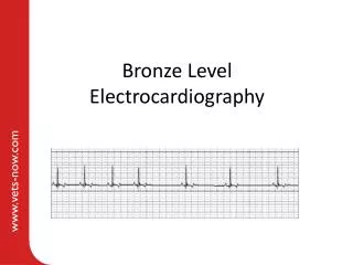
Bronze Level Electrocardiography. Aims. Brief summary of relevant clinical electrophysiology Indications for taking an electrocardiogram (ECG) How to obtain a diagnostic ECG All Covered in Part 1 Basic ECG interpretation. Section 4 – basic ECG interpretation. Definitions. QRS.

E N D
Aims • Brief summary of relevant clinical electrophysiology • Indications for taking an electrocardiogram (ECG) • How to obtain a diagnostic ECG All Covered in Part 1 • Basic ECG interpretation
Definitions QRS R-R interval P T PQ interval
Interpretation – instantaneous heart rate 1 • Use R-R interval printed at bottom of trace (in milliseconds) • There are 60 x 1000 milliseconds in one minute • Heart rate = 60000 / R-R interval(ms) • In this case 60000 / 400 = 150bpm 400ms
Interpretation – instantaneous heart rate 2 • Use a ECG measuring ruler similar to the one shown below • It is important to choose the same scale as the paper speed – usually 25 or 50mm/s • The arrow is placed against the peak of one R wave and the number corresponding to the next R wave shows the heart rate
Interpretation – instantaneous heart rate 2 • Using ruler • Rate here = 150bpm
Interpretation – mean heart rate • Use centimetre ruler • At 25mm/s, 15cm = 6 seconds • No of R waves in 6 seconds x 10 = heart rate in bpm • Rate here = 80bpm 15cm = 6 seconds of trace
If the paper speed is 25mm/s then the information from 1 second of the recording occupies 25mm (=2.5cm) on the paper trace. • Therefore 6 seconds of information will occupy 6 x 25mm = 150mm (=15cm) of the trace. • To convert the number of beats in 6 seconds to the number of beats in 1 minute you multiply by 10 as there are 60 seconds in 1 minute. • Therefore by counting the number of beats within a 6 second period and then multiplying by 10 will give the heart rate in beats per minute (bpm). • In this example there are 8 beats during a 6 second period and therefore 8 x 10 = 80bpm. 15cm = 6 seconds of trace
Normal heart rate • Appropriateness of heart rate depends on species, age, temperament, level of fitness and also concurrent disease especially conditions altering autonomic tone (eg respiratory, gastrointestinal, intracranial) • Dog usually 90-120bpm in consult • Cat usually 140-160bpm in consult • Neonates usually have higher heart rates
Heart rhythm – regular or irregular? • The ratio of P waves to QRS complexes should be 1:1 • What is rhythm and also the P:QRS on traces below: The first 3 complexes on the trace show a normal P-QRS morphology and the rhythm is regular. The 4th beat occurs after a shorter time than the usual R-R interval and therefore is termed a premature beat. This beat (and also the 7th, 10th and 13th beats) have a narrow QRS complex without an obvious P wave. These beats have a similar QRS morphology to the sinus beats suggesting that they are conducted through the ventricles in a similar way to sinus beats and therefore originate proximal to the atrioventricular node. These beats are called supraventricular premature beats often abbreviated to SPCs. P:QRS = 1:1. The rhythm is regularly irregular . This trace shows sinus arrhythmia. P: QRS = 1:1. The rhythm is regular. This trace shows sinus rhythm.
Normal rhythms – sinus arrhythmia Can be seen in normal dogs at rest. The variations in heart rate are caused by rhythmic fluctuations in vagal tone and therefore this rhythm is abolished by sympathetic stimulation (eg stress, fear, pain). In cats in a clinic, if sinus arrhythmia is present then consider conditions altering autonomic tone especially gastrointestinal, respiratory and intracranial disease.