Download
1 / 53
540 likes | 724 Views
This course focuses on optimizing radiation protection in radiography to ensure diagnostic images are obtained with minimal patient exposure. Topics cover technical aspects, equipment, technique, and interpretation for conventional and digital imaging. Radiographers learn how to optimize screen-film systems, film processing, dark room conditions, viewing box characteristics, and radiation protection aspects. Emphasis is placed on maintaining quality by adhering to proper film processing requirements and dark room conditions. Furthermore, the course highlights the importance of skin-focus distance, operator awareness, and procedural protocols to ensure patient safety during radiographic examinations.

E N D
IAEA Regional Training Course on Radiation Protection of patients for Radiographers, Accra, Ghana, 11-15 July 2011 Optimization of protection in radiography – What radiographers can do?
What is optimization in radiography? • To obtain diagnostic images • containing required diagnostic information • with the minimum patient exposure • within prevailing resource limitations 15.1: Optimization of protection in radiography: technical aspects
How to optimize? • Which aspects to consider ? • Equipment • Examination/technique • Interpretation 15.1: Optimization of protection in radiography: technical aspects
Outline • Optimization in conventional radiography - Technical aspects - Radiation protection aspects • Optimization in digital imaging What radiographers can do in optimization
Optimization in Conventional Radiography What radiographers can do in optimization
Film, fluorescent screen or image intensifier Scattered radiation « Latent » radiological image Bone X Soft tissue Air Primary collimation Antiscatter Grid Beam intensity at detector level Image formation in film-screen imaging What radiographers do in optimization
1. Screen film system: A particular intensifying screen used with a particular type of film • Screen-film combination should be selected according to manufacturer’s recommendations • Identification of screen by type and format • type mismatch (use of different types of screens) FOR THE SAME FORMAT is not ADVISABLE • Screen film contact • loss of spatial resolution, blurred image • Cleanliness What radiographers can do in optimization
2. Film processing: automatic processors • Constant temperature • Constant processing time • Automatic replenishment of chemicals • Drying of films BUT • Can introduce artifacts What radiographers can do in optimization
Film processor QC • Most important QC features: • proper film storage • cassette and screen care • processor chemical care • Identify artifacts • processor cleanliness What radiographers can do in optimization
Manual Processing • There are many places where X Ray films are processed manually, in open tanks • Manual processing can be very effective, BUT there can be many quality problems What radiographers can do in optimization
Dark room conditions in some hospitals What radiographers can do in optimization
Basic film processing Requirements • Temperature - constant and optimum • Time - measured • Developer activity (chemical condition) - fresh and unoxidised What radiographers can do in optimization
Dark room Darkroom characteristics • Safelight • number (as low as possible), distance from the table • type and colours of filters • bulb color (red) or adapted to film • External light tightness • Hygrometry (30 - 60%) • Room temperature < 20° • Film storage conditions 15.1: Optimization of protection in radiography: technical aspects
3. Viewing box characteristics Since the viewing conditions are essential for a good interpretation of the diagnostic images, the viewing conditions must be optimal • Cleanliness of external/internal surface • Brightness • homogeneity within the same viewing box • Coloring • color mismatch must be avoided • Environment • ambient light level: 50 lux maximum 15.1: Optimization of protection in radiography: technical aspects
5 7 0 0 5 8 1 0 3 5 1 0 3 8 7 0 6 2 0 0 4 1 6 0 5 9 2 0 5 8 6 0 2 1 5 0 3 1 1 0 Viewing box color and brightness BLUE COLOR WHITE COLOR WRONG CONFIGURATIONS (cd/m2) What radiographers can do in optimization
Example of poor viewing box What radiographers can do in optimization
Radiation protection aspects • Optimization of patient radiation protection requires periodic evaluation of doses and image quality. • Operators of the X Ray system should be aware of the interdependence between technical parameters, dose and image quality • Procedures should be established for each examination to ensure a proper use of equipment. What radiographers can do in optimization
1. Skin focus distance • In general radiography (except dental), the skin-focus distance should not be lower than 30 cm • In radiography with fixedequipment, the skin-focus distance should not be lower than 45 cm • Usually chest x-ray examinations ( chest stand; FFD= 180 (140-200) cm • Abdominal x-ray examinations: FFD= 115 (100-150) cm What radiographers can do in optimization
2. Awareness • e.g. when there are suspicions about a malfunctioning of the X Ray unit or of the imaging chain in a room that might affect the protection of the patient (i.e., because the images become over or underexposed without changing the technique), this should be reported to the responsible person of the room in order to reevaluate the equipment if necessary and to introduce corrective actions. What radiographers can do in optimization
3. Procedures • Image annotation (patient ID, exam date, markers ) on film • Patient instructions and patient positioning • The usual radiological technique for each projection on a given equipment, with details of the associated image device should be written and be readily accessible, close to the control panel. It should be specified for patients of standard as well as uncommon size What can radiographers do in optimization
………Procedures • It is advisable to use the highest kVp (and the lowest mAs) compatible with the image that one expects to obtain. In this way the patient irradiation will be lower, although it might get lower image contrast. Therefore the optimization is to find the proper balance between contrast and dose • The shortest exposure time as possible should be selected, above all when examining non- collaborating patients (shorter time means reduction in kinetic blurring) What can radiographers do in optimization
…. Procedures: x-ray beam limitation • The smallest film and cassette size compatible with the expected image must be used together with automatic collimation. Otherwise the patient would be over-irradiated, by receiving radiation over a larger volume and transversal surface, and the raise in scattered radiation would reduce the image quality and increase the operating personnel doses. • When using equipment without automatic X Ray beam collimation, it should be verified that the radiation field is reduced up to the smallest size compatible with the required image. What can radiographers do in optimization
Periodic evaluation of the image quality • A periodic evaluation (at least once a year) of the image quality obtained in each room allows thedetection of malfunctioning in the imaging chain or in the generator (almost always associated to high patient doses and poor image quality) • A periodic evaluation of the number of rejected and repeated radiographs and the analysis of the causes of rejects and retakes allow the detection of failures in the X Ray equipment, associated image devices, as well as in the examination procedures and in the skill and training of the staff What can radiographers do in optimization
Corrective actions • When some corrective measures are proposed after the detection of a malfunctioning, recording the follow up of their implementation is advisable (in a logbook) What can radiographers do in optimization
Optimization in Digital imaging What can radiographers do in optimization
Radiation dose in digital radiology • Conventional films allow to detect mistakes if a wrong radiographic technique is used: images are too white or too black • Digital technology provides user always with a “good image” since its dynamic range compensates for wrong settings even if the dose is higher than necessary What radiographers can do in optimization
Underexposure results in a “too noisy” image • Overexposure yields good images with unnecessary high dose to the patient Exposure level 2,98 Exposure level 2,36 What radiographers can do in optimization
An underexposed image is “too noisy” Exposure level 1,15 Exposure level 1,87 20: Digital Radiology
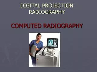
Three approaches to digital radiography • Translate developed film into digital form • (Digitizing conventional films). • II. Capture the radiographic projection by non- • photographic method and digitize during development • (Computed radiography, CR) • Capture the radiographic projection or its fluorescence • directly in digital form (Direct radiography, DR). 15.1: Optimization of protection in radiography: technical aspects
Typical CR system 15.1: Optimization of protection in radiography: technical aspects
Typical DR system 15.1: Optimization of protection in radiography: technical aspects
How to move from film-screen to digital? • Typically the initial setting for the automatic exposure control using CR, should be the same or similar to the existing with film-screen. Optimisation (including possible kV changes) and potential dose reduction should be initiated later once radiologists become familiar with digital. • Audit DICOM header. It contains a lot of useful information. Radiation Protection in Digital Radiology L01 Fundamentals of Digital Radiography
What can a radiographer do? Generally as for film screen imaging • Image annotation • Patient positioning and collimation • Radiographic exposure per examination • Image viewing conditions • Patient dose and image quality evaluation 15.1: Optimization of protection in radiography: technical aspects
Specific for digital imaging • Exposure parameters and image processing (follow • manufacturer guidance • Lower mAs than in film–screen • High or low kVp? (generally lower than in film-screen • protective shielding (assure that area being examined is not covered) • Post processing (adequate, avoid too much processing since artifacts can result) • Image viewing conditions (calibrated to fit purpose) • Cleanliness (extremely important-to avoid artifacts) What radiographers can do in optimization
Improper collimation can cause an incorrect exposure indicator value: Fuji CR S = 578, L=1.8 S = 183, L = 1.9
As seen by radiologist Actual radiation field
Incorrect collimation can cause inappropriate image processing: Agfa CR Automatic processing Manual reprocessing Radiation Protection in Digital Radiology L06 Avoiding Artefacts in Computed Radiography
Processed Unprocessed Gonadal shielding has drawbacks • Ovary locations vary • Shield may obscure clinical features • Shield may interfere with automatic image processing
Image quality and dose • Diagnostic information content in digital radiology is generally higher than in conventional radiology if equivalent dose parameters are used • The wider dynamic range of the digital detectors and the capabilities of post processing allow to obtain more information from the radiographic images What radiographers can do in optimization
Exposure level • Some digital systems provide the user with a so called “exposure level” index which expresses the dose level received at the digital detector and orientates the operator about the goodness of the radiographic technique used • The relation between dose and exposure level is usually logarithmic: doubling the dose to the detector, will increase the “exposure level” to a factor of 0.3 = log(2). What radiographers can do in optimization
Difficulty to audit the number of images per procedure • Deleting useless imagesbefore sending them to the PACS is also very easyin digital fluoroscopy • This makes difficult any auditing of the dose imparted to the patient • The same applies to projection radiography to audit the retakes. What radiographers can do in optimization
Actions that can influence image quality and patient doses in digital radiology (2) • Eliminate post-processing problems, digitizer problems, local hard disk, fault in electrical power supply, network problems during image archiving etc. • Avoid loss of images in the network or in the PACS due to bad identification or others • Reduce artifacts due to incorrect digital post-processing (creation of false lesions or pathologies) What radiographers can do in optimization
Diagnostic reference levels • In digital radiology, the evaluation of patient doses should be performed more frequently than in conventional radiology: • Easy improvement of image quality • Unknown use of high dose technique • Re-assessment of local reference levels when new digital techniques are introduced is recommended to demonstrate the optimization of the systems and to establish a baseline value useful for future patient dose assessment
Artefacts Mispositioning Over-collimation Patient motion Double exposure Inadequate inspiration Overexposed - too dark Underexposed - too light Marker missing or wrong Wrong exam Wrong patient Film lost in processor Conventional Reason for Repeated Exam Radiation Protection in Digital Radiology L03 Optimisation in CR and DR
Artefacts Mispositioning Over-collimation Patient motion Double exposure Inadequate inspiration Overexposed - high exposure index Underexposed - low exposure index Marker missing or wrong Wrong exam Wrong patient Lost image corrupt data, cannot transfer deleted by operator (waste bin) CR/DR Reason for Repeated Exam Radiation Protection in Digital Radiology L03 Optimisation in CR and DR
Remember digital imaging can lead to many artefacts • PSP Plates • Scratches in the plate. • Abdomen of a 3rd trimester pregnant woman.
CR Imaging plates do not last forever Imaging plate artefact Agfa plate defect Same defect, different orientation Artefact remedy: The Imaging plate (IP) must be replaced when cracks occur in clinically useful areas
Imaging plate artefact • PSP Plates • Artifact due to humidity
Imaging plate artefact Phosphor plate misused. Impossible to clean.
Dust on collection optics causes line in slow-scan dimension (Fuji CR) • Dirt on the light guide in the CR reader. The light guide collects light emitted from the imaging plate when it is scanned by the laser. • Artefact remedy: the light guide of the photomultiplier tube was cleaned by service personnel. Radiation Protection in Digital Radiology L06 Avoiding Artefacts in Computed Radiography