Download
1 / 1
20 likes | 118 Views
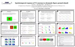
Spatiotemporal response of V1 neurons to chromatic figure ground stimuli Lei Liu 2 , Xiaogang Yan 2,3 , Tai Sing Lee 1,2 Computer Science Department, 2. Center for the Neural Basis of Cognition, Carnegie Mellon University, Pittsburgh, U.S.A.

E N D
Spatiotemporal response of V1 neurons to chromatic figure ground stimuli Lei Liu2, Xiaogang Yan2,3 , Tai Sing Lee1,2 Computer Science Department, 2. Center for the Neural Basis of Cognition, Carnegie Mellon University, Pittsburgh, U.S.A. 3. Center for Vision Research, York University, Canada. Presented by Ryan Kelly and Matt Smith 7 4 10 1 Key Findings: Introduction Magnitude of the figural enhancement effect (within-stimulus comparison, monkey Bo population average) Responses to surround stimulation Strong responses at chromatic border. Significant responses in solid color regions inside figure and outside figure in some neurons, and particularly in multi-unit activities. Responses inside figure stronger than responses outside figure, in both cross-stimuli comparison (RF color same), and within-stimulus comparison (RF colors different). Within-stimulus comparison shows later response is stronger in the figure than in the background regardless of the color preference of the cells (also see panel 7) Population PSTHs (19 cells) from monkey D in response to disc made visible by surround update. 0 msec is the time of surround update. When we see a red square in a green background, the square can be considered as a figure. Since there is no oriented feature inside the receptive field of the neurons, how do V1 neurons respond to the region inside the figure over time? What is the origin of this response? Response to red figure green background stimuli: Response to green figure red background stimuli: Response to red figure compared to response to red ground (cross-stimuli comparison) Response to green figure compared to responses to red ground (within stimulus comparison) Response to blue figure yellow background stimuli: Response to yellow figure blue background stimuli. Receptive field 5 2 8 11 Magnitude of the figural enhancement effect (cross-stimuli comparison of MU population response of monkey Bo) Spatiotemporal responses to color stimuli Distribution of response onset Statistics of the response to surround stimulation Experimental paradigm: Monkeys performed the fixation task. In each trial, the stimuli was presented statically at a location on the screen relative to the receptive field of the neuron (indicated by each cross). Onset of response to color onset (figure) Histograms of onset time of “figural enhancement” as a function of size. Histograms of “figural enhancement” as a function of size. Red Figure versus Red Ground Green Figure versus Green Ground Onset of response to color onset (ground) Details: color is equiluminant (but not cone-isolating). The figure is a 4 by 4 (visual angle) square. Minimally 30 trials were recorded per location. A total of 12 locations per image were examined, with sampling interval = 0.66 degree visual angle. Typical RF size, as mapped by oriented bars, ranged from 0.6 to 1 degree. Recorded eccentricity: 1-3.5 degrees, lower visual fields. 4 monkeys were tested. At least two monkeys were recorded for each test. Temporal responses at each location of three neurons are shown below: Blue Figure versus Blue Ground Yellow Figure versus Yellow Ground Onset of figural enhancement effect 9 6 3 Summary: Chromatic stimuli can elicit significant figural enhancement in V1 neurons. Onset of enhancement response is ~20 ms later than onset time of response to color transition, depending on the color. Sustained responses to color figure are primarily due to the color contrast border in the RF surround. Onset delay of the enhancement response tends to increase as a function of the distance away from the contrast border. Conclusion: Enhancement responses inside the figure arise from propagation of signals from the chromatic contrast border. Statistics of the enhancement effect Dissociating RF and surround stimulation Spatiotemporal responses of three example neurons Distribution of the figural enhancement index (F-G)/(F+G), where F is the response of a single unit or MU within the figure and G is the response of the same single unit or MU within the background: Stimulus display sequence: Monkey D Monkey Bo References Paradigm: The entire screen first changed from gray to the RF color (it could be red, green, yellow or blue). 350 ms later, the RF surround was changed to the opponent color (e.g. red to green), making visible a disc of size 3, 6 or 9 degrees in diameter centered on the RF. The response is compared against the response when the surround was not updated. This paradigm dissociates the response to initial color onset in RF from the response to the disc appearance due to surround update. 1. Lamme VAF. (1995) The neurophysiology of figure-ground segregation in primary visual cortex. J Neurosci. 15:1605-1615. 2. Zipser K, Lamme VAF, Schiller PH. (1996) Contextual modulation in primarl visual cortex. J Neurosci. 16:7376-7389. 3. Lee TS, Mumford D, Romero R, Lamme VAF. (1998) The role of the primary visual cortex in higher level vision. Vision Research. 38:2429-2454. 4. Rossi AF, Desimone R, Ungerleider LG. (2001) Contextual modulation in Primary Visual Cortex of Macaques. J Neurosci. 21:1698-1709. 5. Marcus DS, Van Essen DC. (2002) Scene segmentation and attention in primate cortical areas V1 and V2. J Neurophysiol. 88:2648-2658.