Download
1 / 7
80 likes | 244 Views
Incidental findings on brain MRI. Arrows indicate abnormalities. Vernooij MW et al. (2007) NEJM 357: 1821-1828. Aneurysm of anterior communicating artery (6 mm diameter; proton-density–weighted axial image).

E N D
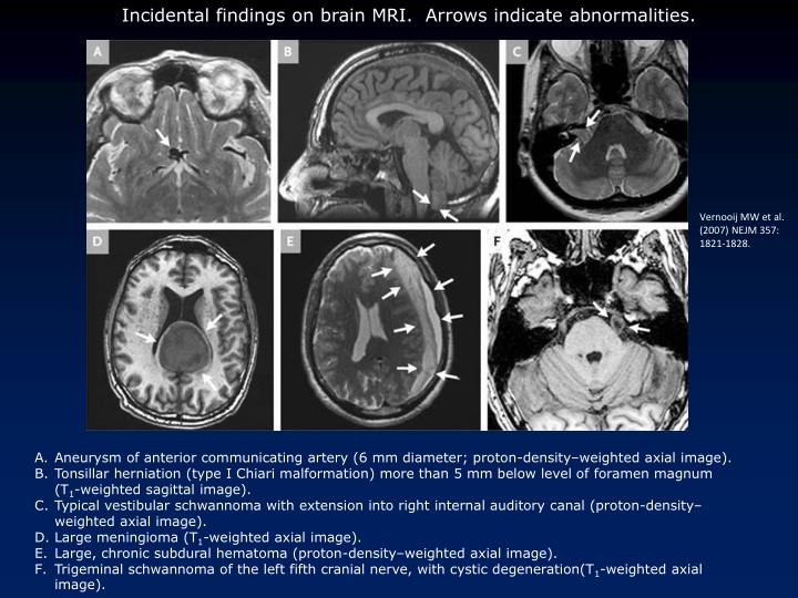
Incidental findings on brain MRI. Arrows indicate abnormalities. Vernooij MW et al. (2007) NEJM 357: 1821-1828. • Aneurysm of anterior communicating artery (6 mm diameter; proton-density–weighted axial image). • Tonsillar herniation (type I Chiari malformation) more than 5 mm below level of foramen magnum (T1-weighted sagittal image). • Typical vestibular schwannoma with extension into right internal auditory canal (proton-density–weighted axial image). • Large meningioma (T1-weighted axial image). • Large, chronic subdural hematoma (proton-density–weighted axial image). • Trigeminal schwannoma of the left fifth cranial nerve, with cystic degeneration(T1-weighted axial image).
Cranial Nerve and Reflex Testing Cranial Nerve Exams http://library.med.utah.edu/neurologicexam/html/cranialnerve_normal.html http://library.med.utah.edu/neurologicexam/html/cranialnerve_abnormal.html Notice: 3,4,6 versions, normal & abnormal1 3,4,6 ductions, abnormal2 Vestibulo-ocular normal (tests 3, 8) 12 normal, abnormal Reflexes Normal Babinski: http://library.med.utah.edu/neurologicexam/html/motor_normal.html#10 Normal Babinski (infant): http://video.google.com/videoplay?docid=-3102473882446365023&pr=goog-sl Positive Babinski (adult): http://www.youtube.com/watch?v=bWKTrUjxkqs 1. Versions: binocular tests, part of a regular exam. Pt. 1 can’t abduct L, i.e. n.6 (L) palsy. Pt.2 limited adduct, elevate, depress L eye, also shows ptosis & dilated pupil, i.e. n.3 (L) palsy. 2. Duction (monocular) tests done iff version results abnormal. Pt can’t medially rotate either eye. Movies from the Neurologic Exam and PediNeurologic Exam websites by Paul D. Larsen, M.D., University of Nebraska Medical Center and Suzanne S. Stensaas, Ph.D., University of Utah School of Medicine. Additional materials for Neurologic Exam are drawn from resources provided by Alejandro Stern, Stern Foundation, Buenos Aires, Argentina; Kathleen Digre, M.D., University of Utah; and Daniel Jacobson, M.D., Marshfield Clinic, Wisconsin.
Anatomical basis of the ear-cough reflex Posterior view of temporal bone 1 vagus nerve, 2 glossopharyngeal nerve, 3 facial nerve, 4 chorda tympani, 5 auricular branch of the vagus (Arnold's nerve), 6 anastomosis with auricular branch of the facial 7, 8 jugular vein, 9 internal carotid artery (reproduced from Tiedemans Z Schr Physiol, 1832) F. Jegoux et al. (2002) Lancet 360: 618
Diagrammatic representation of Arnold's (ear-cough) reflex D. Gupta et al. (1986) Surg Radiol Anat 8 : 217-220
Spontaneous Otogenic PneumocephalusHealthy 54-year-old woman presented with abnormal acoustic sensations, aphasia, visual-field disturbances. She reported no head trauma. See notes. Villa R.A., Capdevila A. N Engl J Med 2008;358:e13, March 20, 2008