Download
1 / 17
170 likes | 330 Views
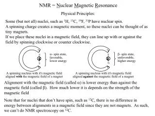
NMR = N uclear M agnetic R esonance. Physical Principles:. Some (but not all) nuclei, such as 1 H, 13 C, 19 F, 31 P have nuclear spin. A spinning charge creates a magnetic moment, so these nuclei can be thought of as tiny magnets.

E N D
NMR = Nuclear Magnetic Resonance Physical Principles: Some (but not all) nuclei, such as 1H, 13C, 19F, 31P have nuclear spin. A spinning charge creates a magnetic moment, so these nuclei can be thought of as tiny magnets. If we place these nuclei in a magnetic field, they can line up with or against the field by spinning clockwise or counter clockwise. Alignment with the magnetic field (called ) is lower energy than against the magnetic field (called ). How much lower it is depends on the strength of the magnetic field Note that for nuclei that don’t have spin, such as 12C, there is no difference in energy between alignments in a magnetic field since they are not magnets. As such, we can’t do NMR spectroscopy on 12C.
NMR: Basic Experimental Principles Imagine placing a molecule, for example, CH4, in a magnetic field. We can probe the energy difference of the - and - state of the protons by irradiating them with EM radiation of just the right energy. In a magnet of 7.05 Tesla, it takes EM radiation of about 300 MHz (radio waves). So, if we bombard the molecule with 300 MHz radio waves, the protons will absorb that energy and we can measure that absorbance. In a magnet of 11.75 Tesla, it takes EM radiation of about 500 MHz (stronger magnet means greater energy difference between the - and - state of the protons) But there’s a problem. If two researchers want to compare their data using magnets of different strengths, they have to adjust for that difference. That’s a pain, so, data is instead reported using the “chemical shift” scale as described on the next slide.
The Chemical Shift (Also Called ) Scale Here’s how it works. We decide on a sample we’ll use to standardize our instruments. We take an NMR of that standard and measure its absorbance frequency. We then measure the frequency of our sample and subtract its frequency from that of the standard. We then then divide by the frequency of the standard. This gives a number called the “chemical shift,” also called d, which does not depend on the magnetic field strength. Why not? Let’s look at two examples. Imagine that we have a magnet where our standard absorbs at 300,000,000 Hz (300 megahertz), and our sample absorbs at 300,000,300 Hz. The difference is 300 Hz, so we take 300/300,000,000 = 1/1,000,000 and call that 1 part per million (or 1 PPM). Now lets examine the same sample in a stronger magnetic field where the reference comes at 500,000,000 Hz, or 500 megahertz. The frequency of our sample will increase proportionally, and will come at 500,000,500 Hz. The difference is now 500 Hz, but we divide by 500,000,000 (500/500,000,000 = 1/1,000,000, = 1 PPM). It’s brilliant. Of course, we don’t do any of this, it’s all done automatically by the NMR machine. Even more brilliant.
The Chemical Shift of Different Protons NMR would not be very valuable if all protons absorbed at the same frequency. You’d see a signal that indicates the presence of hydrogens in your sample, but any fool knows there’s hydrogen in organic molecules. What makes it useful is that different protons usually appear at different chemical shifts (d. So, we can distinguish one kind of proton from another. Why do different protons appear at different d? There are several reasons, one of which is shielding. The electrons in a bond shield the nuclei from the magnetic field. So, if there is more electron density around a proton, it sees a slightly lower magnetic field, less electron density means it sees a higher magnetic field: How do the electrons shield the magnetic field? By moving. A moving charge creates a magnetic field, and the field created by the moving electrons opposes the magnetic field of our NMR machine. It’s not a huge effect, but it’s enough to enable us to distinguish between different protons in our sample.
The Hard Part - Interpreting Spectra Learning how an NMR machine works is straightforward. What is less straightforward is learning how to use the data we get from an NMR machine (the spectrum of ethyl acetate is shown below). That’s because each NMR spectrum is a puzzle, and there’s no single fact that you simply have to memorize to solve these spectra. You have to consider lots of pieces of data and come up with a structure that fits all the data. What kinds of data do we get from NMR spectra? For 1H NMR, there are three kinds each of which we will consider each of these separately: Chemical shift data - tells us what kinds of protons we have. Integrals - tells us the ratio of each kind of proton in our sample. 1H - 1H coupling - tells us about protons that are near other protons.
Chemical Shift Data As previously mentioned, different kinds of protons typically come at different chemical shifts. Shown below is a chart of where some common kinds of protons appear in the d scale. Note that most protons appear between 0 and 10 ppm. The reference, tetramethylsilane (TMS) appears at 0 ppm, and aldehydes appear near 10 ppm. There is a page in your lab handout with more precise values for this chart. Note that these are typical values and that there are lots of exceptions!
Integrals Integrals tell us the ratio of each kind of proton. They are lines, the heights of which are proportional to the intensity of the signal. Consider ethyl acetate. There are three kinds of protons in this molecule, the CH3 next to the carbonyl, the CH2 next to the O and the CH3 next to the CH2. The ratio of the signals arising from each of these kinds of protons should be 3 to 2 to 3, respectively. So, if we look at the height of the integrals they should be 3 to 2 to 3. With this information, we can know which is the CH2 signal (it’s the smallest one), but to distinguish the other two, we have to be able to predict their chemical shifts. The chart on the previous page allows us to make that assignment (the CH3 next to the C=O should appear at ~ 2 PPM, while the other CH3 should be at ~ 1 PPM).
1H - 1H Coupling You’ll notice in the spectra that we’ve seen that the signals don’t appear as single lines, sometimes they appear as multiple lines. This is due to 1H - 1H coupling (also called spin-spin splitting or J-coupling). Here’s how it works: Imagine we have a molecule which contains a proton (let’s call it HA) attached to a carbon, and that this carbon is attached to another carbon which also contains a proton (let’s call it HB). It turns out that HA feels the presence of HB. Recall that these protons are tiny little magnets, that can be oriented either with or against the magnetic field of the NMR machine. When the field created by HB reinforces the magnetic field of the NMR machine (B0 ) HA feels a slightly stronger field, but when the field created by HB opposes B0, HA feels a slightly weaker field. So, we see two signals for HA depending on the alignment of HB. The same is true for HB, it can feel either a slightly stronger or weaker field due to HA’s presence. So, rather than see a single line for each of these protons, we see two lines for each.
More 1H - 1H Coupling What happens when there is more than one proton splitting a neighboring proton? We get more lines. Consider the molecule below where we have two protons on one carbon and one proton on another.
Why are There Three Lines for HB? HB feels the splitting of both HA and HA’. So, let’s imagine starting with HB as a single line, then let’s “turn on” the coupling from HA and HA’ one at a time: Because the two lines in the middle overlap, that line is twice as big as the lines on the outside. More neighboring protons leads to more lines as shown on the next slide.
Splitting Patterns with Multiple Neighboring Protons If a proton has n neighboring protons that are equivalent, that proton will be split into n+1 lines. So, if we have four equivalent neighbors, we will have five lines, six equivalent neighbors… well, you can do the math. The lines will not be of equal intensity, rather their intensity will be given by Pascal’s triangle as shown below. We keep emphasizing that this pattern only holds for when the neighboring protons are equivalent. Why is that? The answer is two slides away.
More About Coupling Earlier we said that protons couple to each other because they feel the magnetic field of the neighboring protons. While this is true, the mechanism by which they feel this field is complicated and is beyond the scope of this class (they don’t just feel it through space, it’s transmitted through the electrons in the bonds). It turns out that when two protons appear at the same chemical shift, they do not split each other. So, in EtBr, we have a CH3 next to a CH2, and each proton of the CH3 group is only coupled to the protons of the CH2 group, not the other CH3 protons because all the CH3 protons come at the same chemical shift.
Not all Couplings are Equal When protons couple to each other, they do so with a certain intensity. This is called the “coupling constant.” Coupling constants can vary from 0 Hz (which means that the protons are not coupled, even though they are neighbors) to 16 Hz. Typically, they are around 7 Hz, but many molecules contain coupling constants that vary significantly from that. So, what happens when a molecule contains a proton which is coupled to two different protons with different coupling constants? We get a different pattern as described in the diagram below. So, if the protons are not equivalent, they can have different coupling constants and the resulting pattern will not be a triplet, but a “doublet of doublets.” Sometimes, nonequivalent protons can be on the same carbon as described on the next slide.
Coupling Constants in Alkenes Coupling constants in alkenes can also differ depending on whether the protons are cis or trans to each other. Note that in a terminal alkene (i.e., an alkene at the end of a carbon chain), the cis and trans protons are NOT equivalent. One is on the same side as the substituent, the other is on the opposite side. The coupling of trans protons to each other is typically very large, around 16 Hz, while the coupling of cis protons, while still large, is a little smaller, around 12 Hz. This leads to the pattern shown below, and an example of a molecule with this splitting pattern is shown on the next slide. There are other times when protons on the same carbon are nonequivalent, which we’ll see later.