Cutting-edge Technology for Virtual Colonoscopy: A Game Changer in Medical Imaging
300 likes | 392 Views
Explore the innovative 3D visualization of the colon using CT Pneumocolon. Learn about "Virtual Dissection" method for fast, accurate, and hardware-independent results in colonoscopy. Discover the benefits and challenges in this groundbreaking medical imaging technique.

Cutting-edge Technology for Virtual Colonoscopy: A Game Changer in Medical Imaging
E N D
Presentation Transcript
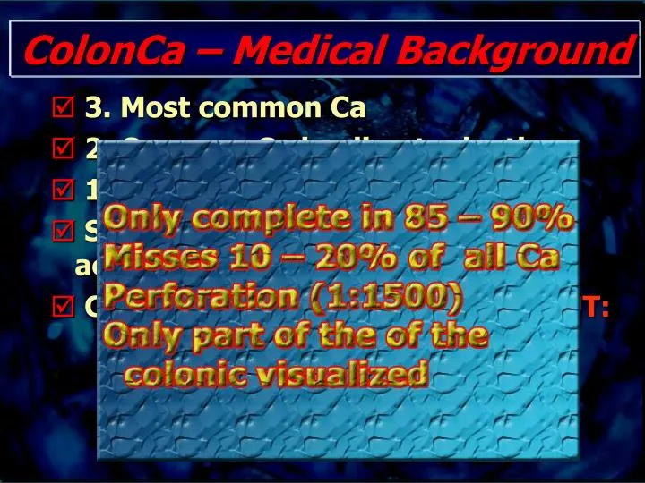
ColonCa – Medical Background • 3. Most common Ca • 2. Common Ca leading to death • 11% of all new cancers/year • Sequence Adenoma – Carcinoma accepted • Colonoskopy = gold standard – BUT:
3D Visualization of the Colon: • CT Pneumocolon • „Interactive Slicing“ • „Fly through“ (CT-colonoscopy) • Virt. Dissection
Problems - General: • Post processing of cross sectional data: • time consuming • hardware intensiv (expensive!) • specialized staff necessary
Problems - CT colonoscopy • Path planning for automated „fly through“ complicated and even operator dependent • Endoscopic view display just a small proportion of the colonic surface -> Ante and - retrograde views necessary
What do you wanna have for CT - colonoscopy? • Inspection of total colonic surface • Fast tool • Minimal interaction • No operator influence • Easy and quick to report • (Hardware independent)
„Virtual dissection“ • Stretch the colon and cut it along it‘s longitudinal axis similiar to the pathologist‘s table
„Virtual dissection“ - How to do? • Data aquisition • Segmentation - Fuzzy connected • Extraction of the centerline (Skeletonisation - Thining) • Calculation of the cross sections • „Remapping“ to 2D
Virt.Diss. - Data Aquisition • MRDCT • Slice thickness 2.5mm • Reconstruction: 1.25mm slice thickness, increment 0.5 - 1.0mm (~600-700 images)
Virt.Diss. - Remapping • Constant Angle Sampling • No distortion in the y direction but adds area distortion. • Can miss objects. • No sense of “size”. • The surface is not sampled uniformally
Virt.Diss. - Remapping • Perimeter Sampling • Surface Sampled uniformally. • No missing elements. If sample step small enough. • Area Preservation. • Deformation in the y direction-Shrinking • Deformation increasing with distance to the vertical center line.
Phantoms - CadavericArtific. Polyps (n=13) Filename: image.691 Table Position: -506.09 Size(Trans, Cor, Sag): 12.9 * 15 * 11.4 Protrusion(Trans, Sag, Cor): 13.8 * 13.8 * 11.2
Phantoms - CadavericArtific. Polyps (n=13) Filename: image.580 Table Position: -450.09 Size(Trans, Cor, Sag): 6.8 * 4.8 * 3.6 Protrusion(Trans, Sag, Cor): 2.6 * 1.6 * 0
Results - Cadaveric Phantom Constant Angle Sampling Perimeter Sampling
Results - Cadaveric Phantom • Polyps appear • as bumps • as asymmetric broadening of folds • Time: • Operator 10min • Total time: 2h
Results - Medical Evaluation • 2 Observer, 13 Polyps: • Sensitivity: 12/13 = 92.3% • Interob. Agreement: 11/13 = 84.6% • Pos.pred.Value: 75-80% • Each Observer overlooked 1 Polyp: • 3.9 * 5.0mm • 3.5 * 2.5mm
Conclusion VirtDiss • Virt. Dissection of the Colon seems to be possible within a reasonable timeframe • Operator interaction minimal (<10min), total time about 2h • Graz enviroment plattform independent • Easy to report, performance excellent • Clinical experience until now limited
Outlook • Using the depth map enhance the image using image processing. • Contour detection. • Curvature detection.
Outlook • „Troubelshhooting Tool“ • „Fecal Tagging“ – for easier patient preparation