The Knee
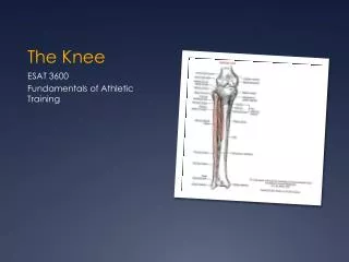
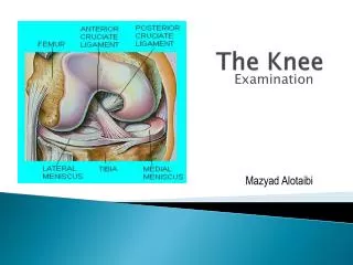
The Knee. Chapter 18. Knee Bony Anatomy. Femur Condyles Lateral Medial Tibia Tibial Plateau Fibula Patella Largest sesamoid in body. Knee Bony Anatomy. Patellofemoral Joint. Point where patella and femur are connected in the trochlear grove. Tibiofemoral Joint.

The Knee
E N D
Presentation Transcript
The Knee Chapter 18
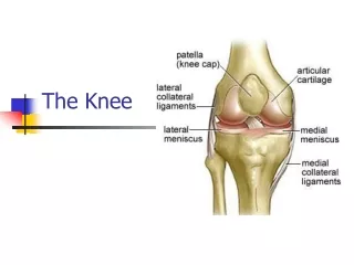
Knee Bony Anatomy • Femur • Condyles • Lateral • Medial • Tibia • Tibial Plateau • Fibula • Patella • Largest sesamoid in body
Patellofemoral Joint • Point where patella and femur are connected in the trochlear grove
Tibiofemoral Joint • Tibia meets with femur • Weight-bearing joint • Hinge joint • Joint capsule • 4 ligaments • Motions: • Flexion • Extension • Rotation of tibia on femur
Patella Malalignment Deviations GenuValgum GenuVarum
Knee Cartilage & Menisci • Articular cartilage • Thin layer of connective tissue over ends of long bones • Lateral & Medial meniscus • Shock absorption • Distribute forces • Improve stability of femur as it rides on tibia • Synovial membrane • Synovial fluid • Lubricates articulating surfaces of joints • Supplies nutrients to articular cartilage
Menisci • Two—medial & lateral • Fibrocartilaginous disks • Act as cushions between ends of femur and tibia/fibula • Top of tibia flat • Condyles of femur rounded • Make knee joint more stable
Menisci • Medial meniscus • C-shaped • Attached to ligaments on back and medial side of knee • Thus does not move freely • And torn more often than lateral • Lateral meniscus • O-shaped • Attached only at back of knee • Moves more freely as knee flex/extend
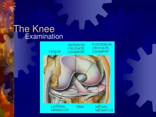
Ligaments of the Knee • Medial Collateral Ligament (MCL) • Lateral Collateral Ligament (LCL) • Anterior Cruciate Ligament (ACL) • Posterior Cruciate Ligament (PCL)
Muscles of the Knee • Quadriceps • Vastus medialis • Vastus intermedius • Vastus lateralis • Rectus femoris • Hamstrings • Biceps femoris • Semitendinosus • Semimembranosus
Quadriceps • Rectus Femoris • Extend knee • Flex hip • Vastus Lateralis • Vastus Medialis • Vastus Intermedius • Extend knee
Hamstrings • Biceps Femoris • Flex knee • Lateral rotate knee • Extend hip • Semitendinosus • Flex knee • Medial rotate knee • Extend hip • Semimembranosus • Flex knee • Medial rotate knee • Extend hip
Muscles of the Knee • Patellar tendon • Sartorius • Flex hip • ER hip • Flex knee • Gracilis • Adduct hip • Flex knee • Pes Ansurine
Gracilis Sartorius
Patellofemoral Problems Signs & Symptoms c/o aching pain in front of knee Gradual onset Pain behind kneecap c/o knee giving way Pain going up stairs Crepitus Pain can increase after prolonged knee flexion
Patellofemoral Problems Causes Treatment Orthotics Muscle strengthening Muscle stretching Patellar tracking taping • Femur internally rotated • Squinting patella • Excessive foot pronation • Lowering of the arch • Thigh hip internal rotators • Weak hip external rotators
Patellar Tendonitis Signs & Symptoms Treatment Modify activity Non-impact activities Stretching quads Ice Specialized bracing & taping • aka Jumper’s knee • Inflammation of the patellar tendon • Anterior knee pain • Local tenderness • Local swelling
Fat Pad Syndrome • Inflammation of infrapatellar fat pad • Fatty tissue lying deep under patellar tendon • Hoffa’s fat pad • Often confused with patellar tendonitis • Pain just below patella • Movement of knee aggravates symptoms • Knee tender to palpation • Swelling in anterior portion of knee • Signs & Symptoms
Fat Pad Syndrome—Treatment • Strengthening exercises • Avoid full knee extension • Leg press • Specialized taping • Ice • NSAIDs
Fat Pad Syndrome—Special Test • Pressure applied to proximal patellar tendon with quadriceps contracted • Stressing only the tendon and not the fat pad • Pressure applied over proximal patellar tendon with relaxed tendon • Allow compression of the fat pad
MCL Sprain MOI Signs & Symptoms Pain & tenderness on medial aspect of knee Joint line Bony attachment sites Limited motion in full flexion and extension Swelling Varying degrees of laxity • Valgus force on medial tibiofemoral joint • Blow to lateral aspect of knee • High-energy twisting maneuver
MCL Sprain—Treatment • PRICE • P: ace, brace, or crutches • Rehab • Submax strengthening in subacute stage, but only if painfree • Bike once gain flex 110-115 degrees • Gentle active & passive stretching • Avoid valgus & twisting forces
LCL Sprain • Not frequently involved in sports injuries • MOI: varus stress on lateral tibiofemoral joint • Signs/symptoms & treatment similar to those of MCL sprain
MCL/LCL Sprain—Grade 1 • Mild tenderness over ligament • Usually no swelling • Pain felt with valgus/varus test but no laxity
MCL/LCL Sprain—Grade 2 • Significant tenderness over ligament • Some swelling seen over ligament • Pain and laxity in joint with stress test, but definite end point
MCL/LCL Sprain—Grade 3 • Complete tear of ligament • Pain can vary • Sometimes not as bad as Grade 2 • When knee stressed, definite joint laxity • Athlete may c/o knee wobbly or unstable
ACL Injuries • Females who participate in soccer and basketball 4-6 times more likely than males who play same sport • 70% are non-contact injuries • Why incidences higher in females?
Female Factors & ACL • Biomechanical factors • Use quads more than hamstrings • Land on flat foot vs toes • Hormonal influences • Estrogen levels • Environmental factors • Anatomic risk factors
ACL Tear • Contact or non-contact • Low to lateral knee • Knee joint in combined position of flexion, valgus, and rotation of tibia on femur • Once stretched or ruptured, will not heal • Often accompanied by meniscus tears and/or MCL sprains
ACL Tear—Signs/Symptoms • Heard or felt “pop” • Rapid effusion • Knee “buckles” or “gives way” • Special testing—Lachman’s or Anterior Drawer • Test’s ligaments integrity • Within first 5 min to avoid protective muscle guarding • Often false-negative testing • F/u with orthopedist • MRI to confirm
ACL Tear—Treatment • Acute: splint, ice, compressive wrap, crutches • Reconstructive surgery necessary to replace ACL • Patellar tendon • Hamstring tendon (Gold standard) • Cadaver • Comprehensive rehab (6 months)