The Knee
The Knee. Anatomy. Mazyad Alotaibi. The Knee Joint Complex. Tibiofemoral Joint* Patellofemoral Joint* Tibiofibular Joint Capsular Pattern – Greater loss of flexion than extension. Bones. Femur – condyles covered with articular cartilage, intercondylar groove, patella groove

The Knee
E N D
Presentation Transcript
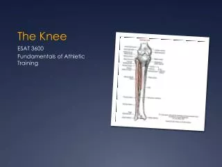
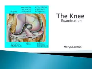
The Knee Anatomy Mazyad Alotaibi
The Knee Joint Complex • Tibiofemoral Joint* • Patellofemoral Joint* • Tibiofibular Joint • Capsular Pattern – Greater loss of flexion than extension
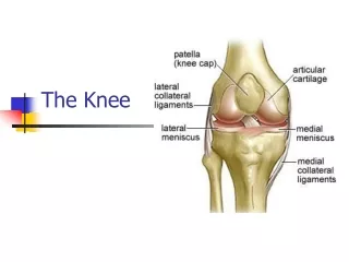
Bones • Femur – condyles covered with articular cartilage, intercondylar groove, patella groove • Tibia – bifid plateau • Patella – medial and lateral articulating facets -increases the lever arm of the quadriceps -increases the distribution of compressive force on the femur in full flexion
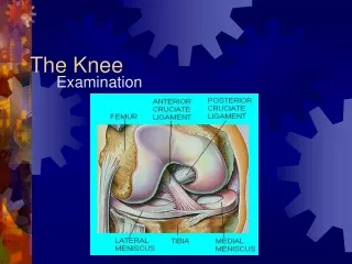
Ligaments Strong ligaments provide the static stability system for the knee
MCL & LCL • Provide medial/ lateral stability and prevent excessive external rotation of the tibia • Frequently injured on the joint line • MCL – Frequently injured by an external rotation strain • Has deep and superficial fibres • LCL – Frequently injured by adduction blow to the knee
ACL • Ant part of intercondylar area • Up, back, lateral • Med asp of lat fem condyle
ACL & PCL • Provide ant / post stability of the knee • ACL – also controls rotation • Commonly injured by forced internal rotation of the femur on a fixed tibia and flexed knee • PCL – strongest -Commonly injured in flexion with an anterior force
Joint capsule – attached to the medial meniscus and MCL • Coronary Ligaments – bind the menisci to the tibia
Menisci • Lateral and medial • Peripherally thicker than central • Transverse lig attaches • Increase stability • Shock absorbing • Lubrication and nutrition • Injured during twisting activities
ITB – anterior and lateral stability and prevent excessive internal rotation of the tibia • Quadriceps – Lat vs med to maintain patella in groove • Sartorius and Gracilis – medial stability, knee and hip flex
Hamstrings • Prevent anterior displacement of the tibia • Pes anserine – sartorius, gracilis and semitendinosis. • Biceps femoris • Primarily produce knee flex and also external rotation of the tibia
Gastrocnemius – primarily ankle flexor also assists with knee flexion • Popliteus – WB external rotation and extension of the tibia • Attaches to the posterior horn of the lateral meniscus and pulls back the posterior horn to unlock the knee
Bursa • Suprapatellar – continuation of the synovial sac • Prepatellar • Deep and superficial infrapatellar • Also semimembranosus and med head of gastroc, • Gastroc heads and capsule • Pes anserine tendon
Surface markings • Joint line • Medial collateral ligament • Lateral collateral ligament • Medial coronarys • Gerdy’s Tubercle • Pes Anseurine Bursa