Knee Joint: Anatomy, Ligaments, and Injuries
450 likes | 503 Views
Explore the complexities of the knee joint, from its bony anatomy to crucial ligaments like ACL and PCL, and common injuries such as patellar dislocation. Learn about stabilizing ligaments, menisci, and muscle functions in maintaining knee stability.

Knee Joint: Anatomy, Ligaments, and Injuries
E N D
Presentation Transcript
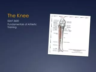
The Knee Some slides adapted from University of Wisconsin Medical School.
The Knee • One of the most complex joints • Provides stability in weight bearing and locomotion • Very vulnerable – especially medially and laterally • Muscles and ligaments provide most of the stability
Instability - Example Patellar dislocation http://www.carletonsportsmed.com/Libraria_medicus/PF_patella_dislocation.JPG
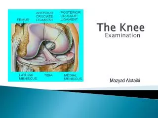
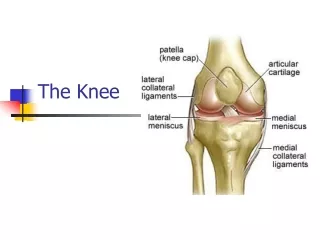
Bony Anatomy – 4 bones Femur Patella Tibia Fibula
Bony Anatomy • Femur: Longest Bone in Body • Tibia: WB bone of lower extremity • Fibula: Site of Muscle Attachment • Patella: Sesamoid Bone • Floating bone • A bone that develops within a tendon
Knee Skeletal Lateral Condyle Head of Fibula Femoral Groove Gerdy’s Tubercle Tibial Tuberosity Pes Anserine
Knee Menisci • 2 oval shaped (semilunar) fibrocartilages • Provides cushion • Avascular(poor blood supply) = decreased healing • Medial – “C” shaped • Lateral – “O” shaped
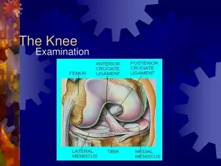
Menisci Medial Meniscus Lateral Meniscus PCL ACL
4 Stabilizing Ligaments Posterior Cruciate • 2 cruciate ligaments • ACL/PCL • 2 collateral ligaments • MCL/LCL Anterior Cruciate Medial Collateral Lateral Collateral
Anterior Cruciate Ligament (ACL) • Extends from tibia posteriorly and laterally to femur • Front of tibia to back of femur • Prevents anterior movement of tibia • Stabilizes against tibial rotation • Main stabilizer
ACL • Torn during cutting motions • Foot planted and knee rotates • More commonly torn in girls • Less muscle, hormones, Q- angle • Surgery • Cadaver graph, patellar tendon, hamstring tendon • About 6-9 months to return to activity
Posterior Cruciate Ligament (PCL) • Extends antiorly and medially from tibia to posterior femur • Prevents tibia from posterior translation • Prevents hyperextension
Medial Collateral LigamentMCL • Medial side • Thick Band of Tissue • Tibia Femur • Resists Valgus Force
Valgus • Outside to Inside Force • MCL resists this force • Occurs in FRONTAL PLANE
Lateral Collateral Ligament LCL • Lateral side • Narrow cord-like band of tissue • Connects femur to • head of fibula • Resists Varus Force
Varus • Inside to Outside Force • LCL resists this force • FRONTAL PLANE
Collateral Ligament Ruptures • 3 degrees of sprains (ligament damage) • Complete tear = 3rd degree sprain
Ruptured Patellar Tendon 3rd degree Strain = muscle/tendon injury
Lab Activity • Partner up • Get a marker • Identify structures of the knee • Patella • Head of fibula • Tibial tuberosity • Pes Anserine • Gerdy’s Tuburcle • MCL • LCL • Medial Joint Line • Lateral Joint Line • Patellar Tendon
Surface Anatomy - Anterior, Extended* Patella Indented Hollow
Surface Anatomy - Anterior, Flexed Patella Tibial Tuberosity Head Of Fibula
Palpation – Anterior* Patella: Lateral and Medial Patellar Facets Superior And Inferior Patellar Facets Medial Fat Pat Lateral Fat Pad Patellar Tendon**
Surface Anatomy - Medial Patella Tibial Tuberosity Medial Femoral Condyle Joint Line Medial Tibial Condyle
Palpation - Medial Medial Collateral Ligament (MCL)* Pes anserine bursa** Medial joint line
Surface Anatomy – Lateral Patella Quadriceps Tibial Tuberosity Head Of Fibula
Palpation – Lateral* Lateral Collateral Ligament (LCL)** Lateral joint line
You should have the following drawn on your partner’s knee • Patella • Head of fibula • Tibial tuberosity • Medial joint line • Lateral joint line • Patellar tendon • MCL • LCL • Pes Anserine • Gerdy’s tubercule
Quadriceps and Patellar Tendons • Quadriceps Tendon • All 4 muscles come together at patella • Patellar Tendon • From inferior patella to tibialtuberosity
Quadriceps • Anterior Thigh Musculature • Four Muscles: • Rectus Femoris • Vastus Lateralis • Vastus Medialis • Vastus Intermedius • Extend the Knee
Rectus Femoris • 2 Joint Muscle • Crosses hip and knee • Flexes Hip • Extend the knee • Converges with rest of quadriceps muscles at tibial tubercle
Hamstrings • Three Muscles • Semimembranosus • Semitendinosus • Biceps Femoris • Common Origin the ischial tuberosity • Flex the Knee