Hearing Things
How Your Brain Works - Week 5 Dr. Jan Schnupp jan.schnupp@dpag.ox.ac.uk HowYourBrainWorks.net. Hearing Things. Sound Signals. Many physical objects emit sounds when they are “excited” (e.g. hit or rubbed).

Hearing Things
E N D
Presentation Transcript
How Your Brain Works - Week 5 Dr. Jan Schnupp jan.schnupp@dpag.ox.ac.uk HowYourBrainWorks.net Hearing Things
Sound Signals • Many physical objects emit sounds when they are “excited” (e.g. hit or rubbed). • Sounds are just pressure waves rippling through the air, but they carry a lot of information about the objects that emitted them.(Example: what are these two objects? Which one is heavier, object A or object B ?) • The sound (or signal) emitted by an object (or system) when hit is known as the impulse response. • Impulse responses of everyday objects can be quite complex, but the sine wave is a fundamental ingredient of these (or any) complex sounds (or signals).
Vibrations of a Spring-Mass System Undamped • F = -k·y (Hooke’s Law) • F = m · a (Newton’s 2nd) • a = dv/dt = d2y/dt2 • -k · y = m · d2y/dt2 • y(t) = yo · cos(t · k/m) Damped 4. –k·y –r dy/dt = d2y/dt2 y(t) = yo·e(-r·t/2m)cos(t·k/m-(r/2m)2) Don’t worry about the formulae! Just remember that mass-spring systems like to vibrate at a rate proportional to the square-root of their “stiffness” and inversely proportional to their weight. https://mustelid.physiol.ox.ac.uk/drupal/?q=acoustics/simple_harmonic_motion
Resonant Cavities • In resonant cavities, “lumps of air” at the entrance/exit of the cavity oscillate under the elastic forces exercised by the air inside the cavity. • The preferred resonance frequency is inversely proportional to the square root of the volume. (Large resonators => deeper sounds).
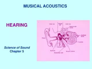
The Ear Organ of Corti Cochela “unrolled” and sectioned
Basilar membrane mechanics:a trade-off between stiffness and inertia
Basilar Membrane Tuning Animation See auditoryneuroscience.com | The Ear
Cochlea Sound Transduction Organ of Corti Sensory “Hairs” (Stereocilia)
Afferent Innervation of the Hair Cells type 1 type 2 Tonotopy
The Outer Hair Cell’s Special Trick https://mustelid.physiol.ox.ac.uk/drupal/?q=ear/dancing_hair_cell
Many Real-World Objects Vibrate at Multiple Frequencies https://mustelid.physiol.ox.ac.uk/drupal/?q=acoustics/modes_of_vibration
The Impulse (or “Click”) • The “ideal click”, or impulse, is an infinitesimally short signal. • The Fourier Transform encourages us to think of this click as an infinite series of sine waves, which have started at the beginning of time, continue until the end of time, and all just happen to pile up at the one moment when the click occurs.
Click Trains & the “30 Hz Transition” • At frequencies up to ca 30 Hz, each click in a click train is perceived as an isolated event. • At frequencies above ca 30 Hz, individual clicks fuse, and one perceives a continuous hum with a strong pitch. sound pressure time https://mustelid.physiol.ox.ac.uk/drupal/?q=pitch/click_train
Harmonic Structure of Click Trains • If we represent each click in a train by its Fourier Transform, then it becomes clear that certain sine components will add (top) while others will cancel (bottom). This results in a strong harmonic structure.
Basilar Membrane Response to Click Trains auditoryneuroscience.com | The Ear
Vocal Folds in Action auditoryneuroscience.com | Vocalizations
Articulators (lips, tongue, jaw, soft palate) move to change resonance properties of the vocal tract. Articulation auditoryneuroscience.com | Vocalizations Launch Spectrogram
Harmonics & Formants of a Vowel Harmonics Formants
As a crude approximation, one might say that it is the job of the ear to produce a spectrogram of the incoming sounds, and that the brain interprets the spectrogram to identify sounds. This figure shows histograms of auditory nerve fibre discharges in response to a speech stimulus. Discharge rates depend on the amount of sound energy near the neuron’scharacteristic frequency. The “Neurogram”
Phase Locking The discharges of cochlear nerve fibres to low-frequency sounds are not random; they occur at particular times (phase locking). Evans (1975) https://mustelid.physiol.ox.ac.uk/drupal/?q=ear/phase_locking
AN Phase Locking to Artificial “Single Formant” Vowel Sounds • Cariani & Delgutte AN recordings Phase locking to Modulator(Envelope) Phase locking to Carrier https://mustelid.physiol.ox.ac.uk/drupal/?q=ear/bm_motion_3
Tonotopicity in the Cochlear Nucleus The base of the BM projects to medial CN, the apex to lateral CN Anteroventral CN Posteroventral CN Anteroventral CN CochlearNerve Dorsal CN Posteroventral CN Cochlea
Neurons of the Cochlear Nucleus Figure 2-16 of Schnupp, Nelken, King “Auditory Neuroscience”
Periodicity Maps in the Midbrain? Neurons in the midbrain or above show much less phase locking to AM than neurons in the brainstem. Transition from a timing to a rate code. Some neurons have bandpassMTFs and exhibit “best modulation frequencies” (BMFs). Topographic maps of BMF may exist within isofrequency laminae of the ICc, (“periodotopy”).
Periodotopic maps via fMRI • Baumann et al Nat Neurosci 2011 described periodotopic maps in monkey IC obtained with fMRI. • They used stimuli from 0.5 Hz (infra-pitch) to 512 Hz (mid-range pitch). • Their sample size is quite small (3 animals – false positive?) • The observed orientation of their periodotopic map (medio-dorsal to latero-ventral for high to low) appears to differ from that described by Schreiner & Langner (1988) in the cat (predimonantly caudal to rostral)
Auditory cortex of Ferret (A), Cat (B) and Macaque Monkey (C) Figure 2-18 of Schnupp, Nelken, King “Auditory Neuroscience”
A pitch area in primates? • In marmoset, Pitch sensitive neurons are most commonly found on the boundary between fields A1 and R. • Fig 2 of Bendor & Wang, Nature 2005
A pitch sensitive neuron in marmoset A1? • Apparently pitch sensitive neurons in marmoset A1. • Fig 1 of Bendor & Wang, Nature 2005
ITD Interaural Time Difference (ITD) Cues ITDs are powerful cues to sound source direction, but they are ambiguous (“cones of confusion”)
ILD at 700 Hz Interaural Level Cues (ILDs) Unlike ITDs, ILDs are highly frequency dependent. At higher sound frequencies ILDs tend to become larger, more complex, and hence potentially more informative. ILD at 11000 Hz
Spectral Cues and the Dorsal Cochlear Nucleus Bushy Multipolar (Stellate) Pyramidal Octopus “Type IV” neurons in the dorsal cochlear nucleus often have inhibitory frequency response areas with excitatory sidebands. This makes them sensitive to “spectral notches” like those seen in spectral localisation cues.
The “Auditory Space Map” in the Superior Colliculus • The SC is involved in directing orienting reflexes and gaze shifts. • Acoustically responsive neurons in rostral SC tend to be tuned to frontal sound source directions, while caudal SC neurons prefer contralateral directions. • Similarly, lateral SC neurons prefer low, medial neurons prefer high sound source elevations.
Where and What Streams in Auditory Cortex? After Romanski et al
Are there “What” and “Where” Streams in Auditory Cortex? Some reports suggest that anterior cortical belt areas may more selective for sound identity and less for sound source location, while caudal belt areas are more location specific. It has been hypothesized that these may be the starting positions for a ventral “what” stream heading for inferotemporal cortex and a dorsal “where” stream which heads for postero-parietal cortex. Figure 2 from Tian & Rauschecker, Science 2001 AnterolateralBelt CaudolateralBelt
Responses to Artificial Vowels Bizley, Walker, Silverman, King & Schnupp - J Neurosci 2009
Joint Sensitivity to Formants and Pitch Vowel type (timbre) Pitch (Hz) Bizley, Walker, Silverman, King & Schnupp - J Neurosci 2009
Azimuth, Pitch and Timbre Sensitivity in Ferret Auditory Cortex JK Bizley, KMM Walker, BW Silverman, AJ King and JWH Schnupp, (2009) Interdependent encoding of pitch, timbre and spatial location in auditory cortex. J Neurosci 29(7):2064-75.