Download
1 / 24
360 likes | 1.06k Views
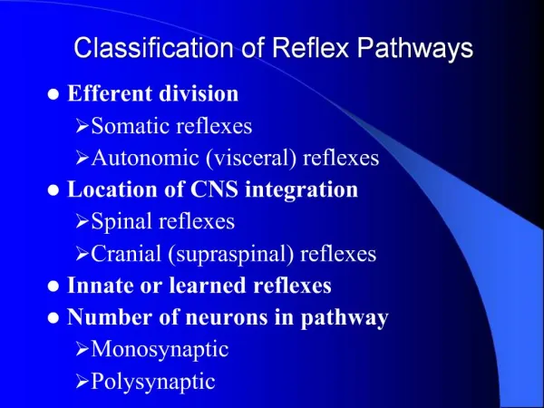
Integrative Physiology I: Control of Body Movement. 13. Neural Reflexes. Somatic Motor Reflexes. Monosynaptic and polysynaptic somatic motor reflexes. Figure 13-1a. Autonomic Reflexes. Sensory neuron. Stimulus. Receptor. CNS integrating center.

E N D
Somatic Motor Reflexes Monosynaptic and polysynaptic somatic motor reflexes Figure 13-1a
Autonomic Reflexes Sensory neuron Stimulus Receptor CNS integrating center All autonomic reflexes are polysynaptic, with at least one synapse in the CNS and another in the autonomic ganglion. Preganglionic autonomic neuron Response Postganglionic autonomic neuron Autonomic ganglion Target cell Some visceral reflexes are spinal reflexes Figure 13-2
Skeletal Muscle Reflexes • Proprioceptors are located in skeletal muscle, joint capsules, and ligaments • Proprioceptors carry input sensory neurons to CNS • CNS integrates input signal • Somatic motor neurons carry output signal • Alpha motor neurons • Effectors are contractile skeletal muscle fibers • Examples of proprioceptors • Muscle spindle • Golgi tendon organ • Joint receptors • Are found in capsules and ligaments around joints
Proprioceptors Muscle spindles are sensory receptors in muscle that control muscle tone and prevent injury from overstretching of the muscle. They are found in all muscles and are tonically active, firing increases as the muscle stretches. Figure 13-3a–b
Muscle Spindles (a) 1 Extrafusal muscle fibers at resting length 1 Spinal cord 3 2 Sensory neuron is tonically active. Sensory neuron endings 2 Sensory neuron Intrafusal fibers of muscle spindle 3 Spinal cord integrates function. 4 Alpha motor neurons to extrafusal fibers receive tonic input from muscle spindles. 4 Alpha motor neuron 5 5 Extrafusal fibers maintain a certain level of tension even at rest. Muscle spindles monitor muscle length and prevent overstretching of the same muscle. The tonic signaling produces muscle tone. Figure 13-4a
Muscle Spindles During a stretch reflex increased firing by the sensory neuron increases signaling by the alpha motor neuron causing the muscle to contract. Figure 13-4b
Alpha-Gamma Coactivation*** (a) If gamma motor axons are cut, the spindle loses activity when muscle contracts. 1 1 Alpha motor neuron fires. Muscle shortens Muscle length 3 2 Muscle contracts. 2 Less stretch on intrafusal fibers 4 Action potential Action potentials of spindle sensory neuron 3 Stretch on center of intrafusal fibers is reduced. Muscle shortens Firing rate of spindle sensory neuron decreases. 4 Time Gamma motor neurons intervate the ends of intrafusal fibers in muscle spindles and keep the sensory neuron active even when the muscle contracts Figure 13-5a
Alpha-Gamma Coactivation (b) Alpha-gamma coactivation maintains spindle function when muscle contracts. 1 1 Alpha motor neuron fires and gamma motor neuron fires. 1 2 Muscle shortens Muscle length 2 Muscle contracts. 3 Intrafusal fibers do not slacken, so firing rate remains constant. 2 Action potentials of spindle sensory neuron 3 Stretch on centers of intrafusal fibers unchanged. Firing rate of afferent neuron remains constant. Muscle shortens 1 Time When an alpha motor neuron fires the muscle contracts and shortens but the gamma motor neuron will also fire and thus there will be stretching at the spindle fiber to keep the tonic firing. Figure 13-5b, steps 1–3
Proprioceptors Golgi tendon organs are sensory receptors in muscle that respond to tension changes in the muscle and attempt to prevent injury from excessively strong contractions. When golgi sensory neuron fibers the efferent signal in inhibitory and thus there is a loss in contraction strength Figure 13-3a, c
Patellar Tendon (Knee Jerk) Reflex Afferent path: Action potential travels through sensory neuron. Receptor: Muscle spindle stretches and fires. Stimulus: Tap to tendon stretches muscle. The patellar tendon (knee jerk) reflex illustrates a monosynaptic stretch reflex and reciprocal inhibition of the antagonistic muscle. Figure 13-7
Patellar Tendon (Knee Jerk) Reflex Afferent path: Action potential travels through sensory neuron. Receptor: Muscle spindle stretches and fires. Integrating center: Sensory neuron synapses in spinal cord. Stimulus: Tap to tendon stretches muscle. The patellar tendon (knee jerk) reflex illustrates a monosynaptic stretch reflex and reciprocal inhibition of the antagonistic muscle. Efferent path 1: Somatic motor neuron onto Efferent path 2: Interneuron inhibiting somatic motor neuron Figure 13-7
Patellar Tendon (Knee Jerk) Reflex Afferent path: Action potential travels through sensory neuron. Receptor: Muscle spindle stretches and fires. Integrating center: Sensory neuron synapses in spinal cord. Stimulus: Tap to tendon stretches muscle. The patellar tendon (knee jerk) reflex illustrates a monosynaptic stretch reflex and reciprocal inhibition of the antagonistic muscle. Efferent path 1: Somatic motor neuron onto Effector 1: Quadriceps muscle Efferent path 2: Interneuron inhibiting somatic motor neuron Response: Quadriceps contracts, swinging lower leg forward. Effector 2: Hamstring muscle Response: Hamstring stays relaxed, allowing extension of leg (reciprocal inhibition). Figure 13-7
Flexion Reflex and the Crossed Extensor Reflex Ascending pathways to brain Spinal cord 3a Gray matter 2 Spinal cord Sensory neuron White matter Painful stimulus activates nociceptor. 1 Primary sensory neuron enters spinal cord and diverges. 2 Nociceptor Painful stimulus 1 3a One collateral activates ascending pathways for sensation (pain) and postural adjustment (shift in center of gravity). Figure 13-8, steps 1–3a
Flexion Reflex and the Crossed Extensor Reflex Ascending pathways to brain Spinal cord 3a Gray matter 2 Spinal cord Sensory neuron White matter Painful stimulus activates nociceptor. 1 3b Primary sensory neuron enters spinal cord and diverges. 2 Nociceptor Alpha motor neurons Painful stimulus 1 Extensors inhibited 3a One collateral activates ascending pathways for sensation (pain) and postural adjustment (shift in center of gravity). Flexors contract, moving foot away from painful stimulus. Withdrawal reflex pulls foot away from painful stimulus. 3b Figure 13-8, steps 1–3b
Flexion Reflex and the Crossed Extensor Reflex Ascending pathways to brain Spinal cord 3a Gray matter 2 Spinal cord Sensory neuron White matter Painful stimulus activates nociceptor. 1 3c 3b Primary sensory neuron enters spinal cord and diverges. 2 Nociceptor Alpha motor neurons Painful stimulus 1 Extensors inhibited 3a One collateral activates ascending pathways for sensation (pain) and postural adjustment (shift in center of gravity). Extensors contract as weight shifts to left leg. Flexors contract, moving foot away from painful stimulus. Withdrawal reflex pulls foot away from painful stimulus. 3b Flexors inhibited Crossed extensor reflex supports body as weight shifts away from painful stimulus. 3c Figure 13-8, steps 1–3c
Movement Classification Three types of movement: Reflex (simplest) voluntary (most complex) Rhythmic (a combination of relex and voluntary).
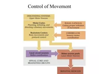
CNS Integrates Movement • Spinal cord integrates spinal reflexes and contains central pattern generators • Brain stem and cerebellum control postural reflexes and hand and eye movements • Cerebral cortex and basal ganglia • Voluntary movement- can become reflexive once well learned Rhythmic movement is initiated at the cerebrum but maintained by interneurons in the spinal cord
Integration of Muscle Reflexes Reflexes are managed by the spinal cord, cerebellum, and brain stem. They do not require input from the cerebrum. However, sensory input is send to the cerebrum so we aware of what happens. Figure 13-9
CNS Control of Voluntary Movement Voluntary movement can be planned based on postural reflex information. There are 3 phases, sensory feed back is used in the first two. Figure 13-10
Voluntary Movement Feedforward reflexes and feedback of information during movement Figure 13-13
Visceral Movement • Contraction of cardiac and smooth muscle • Moves material in hollow organs by changing the shape of the organ • Controlled by ANS as a reflex • Some create own action potentials • Muscle can respond to hormones or signaling from neighboring cells through gap junction