Download
1 / 19
240 likes | 519 Views
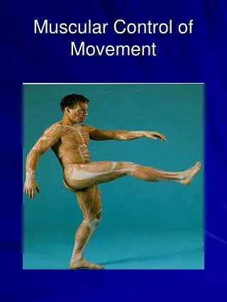
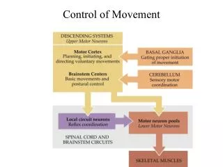
Control of Body Movement. A General Scheme For Motor System Organization. 1. Major Motor Pathways. Neuronal Reflexes. The integration of sensory information into a involuntary response is the hallmark of a reflex .

E N D
1 Major Motor Pathways
Neuronal Reflexes • The integration of sensory information into a involuntary response is the hallmark of a reflex. • All neuronal reflex begin with the activation of sensory receptors by a stimulus.
Reflexes Require Feedback • In a negative feedback pathway, the response of a system removes or opposes the stimulus signal in order to keep the system at or near a set point. -ve feedback keeps the CNS informed about the state of sensory receptors. • Some reflexes have feedfoward component which allows the body to anticipate the stimulus and begin the response. In +ve feedback loops, the response reinforces the stimulus rather than remove or decrease it. Eg. contraction of the uterus during childbirth.
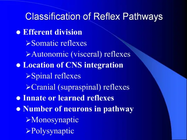
Classification of Neuronal Reflexes • Reflex pathways consist of an interconnected network of neurons which link sensory receptors to muscles and glands. They can be classified in the following ways:
Monosynaptic and Polysynaptic Reflex • Monosynaptic reflex formed by two neurons forming a single synapse. • Most reflexes are polysynaptic, consisting of three or more neurons.
Sensory Receptors in Skeletal Muscle • Skeletal muscle reflexes are involved in almost everything we do. • Sensory inputs are required to control these reflexes. • Motor neurons (MNs) to skeletal muscles are excitatory, and when activated, cause muscles to contract. If the muscles need to be relaxed, the CNS needs to inhibit MN activity. • Sensory receptors in muscles sense changes in tension and length, and activate muscle reflexes. • Three types of sensory receptors in muscle: a) muscle spindles; b) Golgi tendon organs; c) joint capsule mechanoreceptors (proprioceptors). • These three receptors send information to the CNS about the relative positioning of bones linked by flexible joints.
Two Different Types of Motor Neurons Carry Information to Muscle Following Sensory Information Processing • Once sensory information is processed in the CNS, it is sent back to muscles by two different types of motor nurons. • Alpha motor neurons (-MNs): Innervate normal skeletal muscle fibres (also known as extrafusal muscle fibres). APs in these neurons produce muscle contraction. • Gamma motor neurons (-MNs): are smaller motor neurons associated with specialized muscle fibres within sensory receptors.
Muscle Spindles Respond to Stretch • Muscle spindles send information about muscle length to the CNS. • They are small elongated structures arranged in parallel along extrafusal muscle fibres. They are scattered throughout the muscle. • Each spindle consists of connective tissue ensheathing a group of intrafusal muscle fibres. • Intrafusal fibres are modified fibres which lack myofibrils at the centre, but contain contractile ends. They contract when -MNs fire. • Sensory neurons wrap around the middle of the fibres and project to the spinal cord. They fire when the ends of the intrafusal fibres stretch.
Muscles Maintain Muscle Tone • When muscles are at resting length, the spindles are slightly stretched causing sensory neurons to generate tonic activity. As a result of sensory neurons activating -MNs resulting in muscles maintaining a certain level of tension, or tone at rest.
Length of Intrafusal Fibres Affects Sensory Neuron Activity • Muscle stretch also stretches the spindles, causing sensory neurons to fire. • This in turn causes the muscle to contract. • Contraction relieves the stretch on spindles, and reduces sensory neuron AP firing. • The contraction acts as a negative feedback to diminish the reflex. • The reflex pathway in which muscle stretch initiate a contraction is known as a stretch reflex.
-MNs Keep Spindles Functioning During Contraction • Normally, and MNs fire together during muscle contraction. • This causes both intrafusal and extrafusal fibres to contract together. • Contraction of intrafusal fibres causes their middle portion to stretch, causing sensory neurons to fire.
Myotatic Stretch Reflex And Reciprocal Inhibition • The synergistic and antagonistic muscles that control a single joint are known as a myotatic unit. • The simplest reflex in a myotatic unit is a monosynaptic stretch reflex. • The knee jerk reflex is an example of a monosynaptic stretch reflex. • In addition to contraction, there’s relaxation of antagonistic muscles (reciprocal inhibition) upon inhibition of their -MNs from firing.
Summary • Integration of sensory information into involuntary responses is the hallmark of a reflex. Feedback regulates reflexes. • Reflexes can be both monosynaptic and polysynaptic. • Sensory receptors in the muscle send information into the CNS to in turn regulate motor neuron activity. • - and -MNs to muscles are activated following sensory information processing. • Sensory receptors in muscle sense tension and length. • Muscle spindle receptors respond to stretch. Their length is affected by muscle length. • - and -MNs are activated together during muscle contraction. -MNs keep spindle functioning during muscle contraction. • The knee jerk reflex involves contraction of quadriceps and relaxation of hamstrings (reciprocal inhibition). Sensory neurons from quadriceps muscle spindle excite -MNs to the quadraceps, and activate inhibitory neurons which suppress AP firing in -MNs to hamstrings. My Lab Website: http://www.neurobio.arizona.edu/faculty/karunanithi/index.php
References Tortora, G.J. & Grabowski, S.R (2003). Principles of Anatomy & Physiology.New Jersey: John Wiley & Sons. Ch. 13, pp.426-433; Ch.15, pp.505-506; . Silverthorn, D.U (1998). Human Physiology: An Integrated Approach. New Jersey: Prentice Hall. Ch.13, pp.362-374.