Download
1 / 17
200 likes | 568 Views
Neurological Control of Movement. The Structure and Function of the Nervous System. The Neuron. The Cell Body (nucleus) The Dendrites the receivers The Axon the transmitter contains the axon terminals

E N D
Neurological Control of Movement The Structure and Function of the Nervous System
The Neuron • The Cell Body (nucleus) • The Dendrites • the receivers • The Axon • the transmitter • contains the axon terminals • contains the synaptic knobs that release chemicals known as neurotransmitters. • The axon hillock decides if the impulse is a graded potential or an action potential.
The Neuron • Node of ranvier • Myelin sheath • Saltitory conduction: the impulse skips from node to node and is a faster method of impulse travel.
The Nerve Impulse • Nerve Impulse: an electrical charge that passes from one neuron to the next neuron or muscle fiber. • Resting Membrane Potential: the separation of charges across the membrane (polarized). • a constant RMP of -70 mV is the function of the sodium-potassium pump. • Depolarization: when the charge difference decreases (< -70 mV), moving closer to zero (ie -20 mV). • Hyperpolarization: when the charge difference increases (> -70 mV), moving farther from zero (ie -120 mV).
The Nerve Impulse • Graded Potentials: local changes in the neuron membrane to cause an inefficient charge difference. • Action Potentials: a rapid and substantial depolarization (excitation) of the neurons membrane. • axon hillock- measures the summation of impulses and determines the threshold for an action potential • All-Or-None Principle • Sequence of events [3.2]
The Synapse • Synapse: is the site of impulse transmission from one neuron to another neuron or muscle fiber. • axon terminals- release acetylcholine • synaptic cleft • receptors- of a neuromuscular junction at the sarcolemma of a muscle fiber. [3.4]
The Synapse • Excitatory Postsynaptic Potential (EPSP’s) can be either depolarizations (excites) or hyperpolarizations (inhibits) • Inhibitory Postsynaptic Potentials (IPSP’s) are only hyperpolarizations (inhibits)
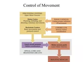
The Brain and Spinal Cord • Cerebrum: site of mind and intellect, motor control, sensory input and interpretation. • Frontal Lobe: general intellect and motor control • Temporal Lobe: auditory input and its interpretation • Parietal Lobe: general sensory input and its interpretation • Occipital Lobe: visual input and its interpretation
The Brain and Spinal Cord • Diencephalon: sensory integration and homeostasis of the body’s internal environment. • Thalamus: interprets sensory input and relays it to the appropriate area of the brain. • Hypothalamus: maintains homeostasis.
The Brain and Spinal Cord • Cerebellum: movement control. • Brain Stem: relays information between the brain and the spinal cord. • Spinal Cord: tracts of nerve fibers that allow two-way conduction of nerve impulses. • afferent -vs- efferent
The Peripheral Nervous System • The PNS contains 12 pairs of cranial nerves and 31 pairs of spinal nerves. • Sensory neurons enter the spinal cord through the dorsal root. • mechanoreceptors (touch) • thermoreceptors (temperature) • nociceptors (pain) • chemoreceptors (oxygen, glucose, electrolytes, etc.) • kinesthetic receptors (movement in joints, balance, etc.) ie. golgi tendon organs
The Peripheral Nervous System • Motor neurons leave the spinal cord through the ventral root. • Create muscle contraction • Create muscle inhibition
The Autonomic Nervous System • The ANS controls your body’s involuntary internal functions. • Sympathetic Nervous System (fight or flight mechanism) • inc. H.R. and cardiac contraction • coronary vessels dilate increasing B.P. & blood flow • bronchodilation, inc. metabolic rate & mental capabilities • glucose is released from the liver into the blood
The Autonomic Nervous System • Parasympathetic Nervous System (housekeeping system) • carry’s out digestion, urination, & life support • conserves energy • decreases blood flow • decreases breathing rate
Sensory Motor Integration • Sensory Motor Integration: is the communication of the sensory and motor nerve pathways. [3.1] • Reflex: when sensory impulses terminate at the spinal cord and are integrated there. • Motor Control: controlled by impulses conducted by motor (efferent) neurons from the brain. • Muscle Spindles: create reflexive muscle contractions of the agonist muscle to resist further stretching. • Golgi Tendon Organs: are sensitive to tension which excite the antagonist muscles to contract.
Muscle Fiber Recruitment • Each muscle fiber is innervated by only one motor neuron, but each motor neuron innervates up to several thousand muscle fibers. • Principle of Orderly Recruitment • Motor units with smaller motor neurons (ST) will be recruited first, larger motor neurons (FTb) last. • Motor units with a smaller number of muscle fibers will be recruited first.