Download
1 / 26
330 likes | 1.28k Views
PHYSIOLOGY OF CONTROL OF BREATHING. Prof. Sultan Ayoub Meo MBBS, M.Phil, Ph.D (Pak), M Med Ed (Dundee), FRCP (London), FRCP (Dublin), FRCP (Glasgow), FRCP (Edinburgh) Professor and Consultant, Department of Physiology, College of Medicine, King Saud University, Riyadh, KSA.

E N D
PHYSIOLOGY OF CONTROL OF BREATHING Prof. Sultan Ayoub Meo MBBS, M.Phil, Ph.D (Pak), M Med Ed (Dundee), FRCP (London), FRCP (Dublin), FRCP (Glasgow), FRCP (Edinburgh) Professor and Consultant, Department of Physiology, College of Medicine, King Saud University, Riyadh, KSA
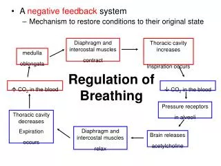
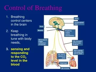
REGULATION OF RESPIRATION Respiration is regulated by three different mechanisms: Nervous regulation Chemical regulation Peripheral chemoreceptor control system.
NERVOUS REGULATION • Dorsal respiratory group • Ventral respiratory group • Pneumotaxic center • Apneustic center
NERVOUS REGULATION Controls automatic breathing. Consists of interacting neurons that fire either during inspiration (I neurons) or expiration (E neurons).
NERVOUS REGULATION • I neurons: Located primarily in dorsal respiratory group (DRG): • Regulate activity of phrenic nerve. • Project to and stimulate spinal interneurons that innervate respiratory muscles. • E neurons: Located in ventral respiratory group (VRG): • Passive process. • Controls motor neurons to the internal intercostal muscles. • E neurons inhibit the I neurons. • Rhythmicity of I and E neurons may be due to pacemaker neurons.
NERVOUS REGULATION Apneustic center: Promote inspiration by stimulating the I neurons in the medulla. Pneumotaxic center: Antagonizes the apneustic center. Inhibits inspiration.
DORSAL RESPIRATORY GROUP OF NEURONS Dorsal respiratory group of neurons are located bilaterally in the dorsal portion of the medulla oblongata in / close to the nucleus of the tractus solitarius. Dorsal group of neuron is made up of I neurons. They receive afferents from the air ways and carotid and aortic bodies which terminate in the nucleus of tractus solitarius.
DORSAL RESPIRATORY GROUP OF NEURONS Functions: On stimulation initiate normal inspiration Rhythmically discharges inspiratory signals Inspiratory signals begin weekly and increase in ramp fashion for 2 seconds, then cease for next 3 seconds and then begin another cycle.
VENTRAL RESPIRATORY GROUP OF NEURONS Ventral respiratory group of neurons extend through the nucleus ambigus and nucleus retroambigus in the ventrolateral part of the medulla oblongata. The ventral group has [E] neurons at its caudal end [I] neurons in its mid portion [E] neurons at its rostral ends. The neurons in the rostral end of this group appear to inhibit [I] neurons during expiration.
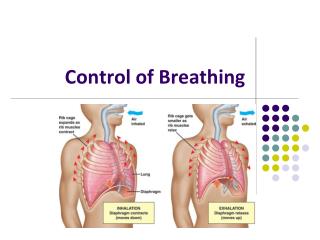
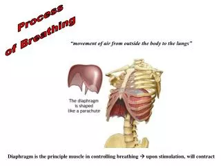
FUNCTIONS OF VENTRAL RESPIRATORY GROUP OF NEURONS Ventral respiratory group neurons are inactive during normal quiet respiration. Normal quiet breathing is caused by repetitive inspiratory signals from the dorsal respiratory group, transmitted mainly to the diaphragm. Expiration results from the elastic recoil of the lungs. These neurons provide active role / strong discharge during forceful expiration.
PNEUMOTAXIC CENTER Pneumotaxic center located dorsally in the nucleus parabrachialis of the upper pons, transmits impulses to the inspiratory area. Functions: Transmit signals to the dorsal inspiratory areas to switch off the inspiratory ramp signals, controlling the duration of the filling phase of the lungs. When these signals are strong inspiration lasts for 0.5 sec. When weak, inspiration lasts as long as 5 seconds, filling the lungs with excess air. Stimulation of the pneumotaxic center limits the period of inspiration, It increases the rate of respiration
APNEUSTIC CENTER Apneustic center: Situated in lower pons. Functions: It send signals to the dorsal respiratory group of neurons to prevents the switch off of inspiratory ramp signals Stimulation of this centre prolongs the period of inspiration. An increase in the duration of inspiration result in a deeper and more prolonged inspiratory effort. The rate of respiration becomes slow because of the greater depth of inspiration
CHEMICAL REGULATION OF RESPIRATION Respiratory system maintain the concentration of CO2 and O2 CO2 is most important stimulus for regulating respiratory rate. Effects of H+ and CO2 on the chemosensitive area: Effects of blood H+ ions:H+ ions that provide the important stimulus for regulating the rate of respiration, blood H+ ions cannot effect the chemosensitive area alone because it cannot cross the blood brain barrier and blood C.S.F barrier. Effects of blood CO2: Blood CO2 can cross the blood brain and blood C.S.F barriers, CO2 in blood combines with H2O to form carbonic acid. This CO2+H2O form H2CO3
CHEMICAL REGULATION OF RESPIRATION Carbonic acid rapidly dissociates into H+ ions and bicarbonate (HCO3-) ions. Increase in CO2 will increase the H+, but on the other hand a decrease in CO2 will cause a decrease in H+ ions. H+ ions stimulate the chemosensitive areas.
Chemoreceptors 2 groups of chemo-receptors that monitor changes in blood PC02, P02, and pH. Central: Medulla. Peripheral: Carotid and aortic bodies. Control breathing indirectly via sensory nerve fibers to the medulla (X, IX).
PERIPHERAL CHEMORECEPTORS Effects of oxygen: The peripheral chemoreceptors detect changes in PO2. The arterial PO2 falls from 104 mm Hg, impulses from these receptors are carried to the brain via the vagus and glossopharyngeal nerves, result in an increased rate and depth of respiration. Effect of decreased pH (increased H+ ions): Increased alveolar ventilation lowers the PCO2 in the arterial blood and reduces the amount of acid, which tends to return the arterial pH to normal.
PERIPHERAL CHEMORECEPTORS Effects of CO2: CO2 stimulates the peripheral chemoreceptors. Peripheral chemoreceptors are stimulated by decreased or increased CO2, increased H+ ion concentration, and decreased pH and low O2. When peripheral chemoreceptors are stimulated, the impulses transmitted from these receptor sites to the dorsal inspiratory area causes the switch off of the inspiratory ramp signals. Since the period of inspiration becomes limited there is an increase in the rate of respiration.
CHEMORECEPTOR CONTROL • Central chemoreceptors: • More sensitive to changes in arterial PC02. • H20 + C02 • H+ cannot cross the blood brain barrier. • C02 can cross the blood brain barrier and will form H2C03. • Lowers pH of CSF. • Directly stimulates central chemoreceptors.
CHEMORECEPTOR CONTROL • Peripheral chemoreceptors: • Are not stimulated directly by changes in arterial PC02. • H20 + C02 H2C03 H+ • Stimulated by rise in [H+] of arterial blood. • Increased [H+] stimulates peripheral chemoreceptors.