Cells
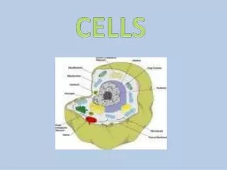
Cells. Chapter 3. I. Overview Cell Membrane Cytoplasm Cytosol Organelles Nonmembranous : Cytoskeleton, Microvilli , Centrioles , Cilia, Flagella, Ribosomes Membranous: Mitochondria, Nucleus, Endoplasmic Reticulum, Golgi Apparatus, Lysosomes , Peroxisomes , Vesicles .

Cells
E N D
Presentation Transcript
Cells Chapter 3
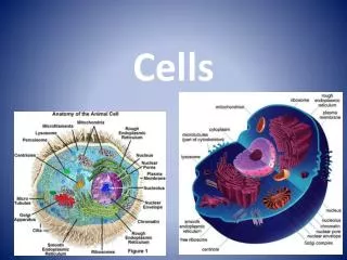
I. Overview • Cell Membrane • Cytoplasm • Cytosol • Organelles • Nonmembranous: Cytoskeleton, Microvilli, Centrioles, Cilia, Flagella, Ribosomes • Membranous: Mitochondria, Nucleus, Endoplasmic Reticulum, Golgi Apparatus, Lysosomes, Peroxisomes, Vesicles
II. Plasma Membrane (Cell Membrane) • "Fluid Mosaic Model" - plasma membrane is composed of a double layer (bilayer) of phospholipid molecules with proteins that float/move among the phospholipids, yet the plasma membrane is stable. • Proteins function.... • As cell markers for recognition by immune system • As receptors (e.g for hormones) • As catalysts • Transportation
Proteins in the membrane... • integral proteins (maintain selective transport) • peripheral proteins (catalyst and mechanical function) • The plasma membrane also contains a myriad of biological compounds such as glycoproteins, glycolipids, and proteoglycans (all referred to as glycocalyx) that extend outward from the plasma membrane
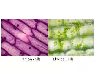
III. Cytoplasm • Cytoplasm is the material found inside the cell and is divided into two subdivisions: cytosol and organelles. • Cytosol (intracellular fluid) contains dissolved nutrients, ions, soluble and insoluble proteins, and waste products. • Organelles are structures that perform specific functions within the cell and are classified as membranous and non-membranous.
IV. Membrane Transport Processes • Transportation of materials across the cell membrane is determined by the components in the membrane that impart permeability. • Most cells have selective permeability, free passage of some materials and restricts the passage of others. • Permeability may be based on size, electrical charge, molecular shape, solubility, etc... Passage across the membrane is classified as active (requiring energy) and passive (not requiring energy)
Membrane transport processes: • Passive • Diffusion - net movement of particles from an area of higher concentration to an area of lower concentration. • Osmosis - diffusion of water through a selectively permeable membrane • Facilitated diffusion - diffusion of a substance with the aid of a membrane carrier. • Filtration - movement of water and solutes through a semipermeable membrane from a region of higher hydrostatic pressure to a region of lower hydrostatic pressure
b) Osmosis – movement of a solvent (water) through a semi- permeable membrane down a concentration gradient (higher to lower) Solutions: Isotonic Hypertonic Hypotonic
A human red blood cell is composed of 0.9% salt and 99.1% water. If this cell is placed in a solution of 0.9% salt and 99.1% water (saline) the solution is isotonic and the blood cell will remain unchanged
The same RBC is placed in a beaker of distilled water (100% H2O and 0% salt), water will enter the cell and cause it to burst (lysis). Water goes from higher conc. to lower conc. This solution is hypotonic (hypo=less salt in solution).
The same RBC is placed in a beaker of 50% salt water (50% H2O and 50% salt), water will leave the cell and cause it to shrink (crenation). Water goes from higher conc. to lower conc. This solution is hypertonic (hyper=more salt in solution).
Active • Active transport - movement of a substance (with the aid of a membrane carrier) through a membrane against its concentration gradient. • Exocytosis- substances enclosed in a vesicle fuses with the plasma membrane, the vesicle then ruptures, releasing the substances outside the cell. • Endocytosis(types): • Phagocytosis - the cell membrane extends outward and encloses large particles which are then transported into the cell. • Pinocytosis - particles attach to the cell membranes which collapses, causing particles to be taken into the cell. • Receptor-mediated - pinocytotic movement initiated by protein receptors on the plasma membrane.
Movement of particle may be.... • Symport - movement of two or more different kinds of material in the same direction across the cell membrane. • Uniport - movement of one type of material in one direction across the cell membrane. • Antiport - moving two types of material across the cell membrane in opposite directions.
V. Cell Division (Cell Life Cycle) • Multicellular organisms develop from a zygote, which is formed by the fusion of a sperm and an egg (gametes). Each gamete has half the compliment of chromosomes (haploid number) and when combined gives rise to a zygote with a complete set (diploid number) of chromosomes. In order for the zygote to develop into a multicellular organism, it must repeatedly undergo cellular divisions. The series of events a cell (or zygote) undergoes that ultimately produces a new cell is called the cell cycle.
Nucleus - located in the center of the cell - controls all functions of organelles - cell reproduction/division takes place - DNA (Deoxyribonucleic Acid) is housed - blueprint of heredity - as cell divides the DNA coil tightly, called chromatin, to form chromosomes (46) - bound by nuclear envelope: double layered membrane enclosing nucleoplasm Nucleoli: are darkly stained areas within the nucleus that indicate rapid RNA synthesis.
Cell Growth and reproduction: produces two identical daughter cells from one parent cell - cell life cycle has two major sections Interphase (cell growth; not dividing) G1phase : growth S phase: growth and DNA synthesis G2phase: growth and final preparation for cell division
Mitotic phase (M): dividing phase Prophase Metaphase Anaphase Telophase IPMAT
Interphase: G1 , S and G2 phases. (90% of its time) G1 (gap 1): cell grows vigorously and metabolically very active. - depending on cell type, this phase may last minutes to years. - centrioles begin replicating S: DNA replicates itself; chromatin condenses. Ensures daughter cells receive identical genetic information.
G2: phase is very brief. - centriole replication is complete - ready to divide
Mitotic phase (M): Prophase: Chromosomes are visible. - early prophase: longest phase of mitosis. - chromatin condenses to form chromosomes. - centriole pairs start to separate/nuclear membrane breaks down. - mitotic spindles (microtubules) start to develop
late prophase: - centrioles migrate away from each to opposite poles of cell.
Metaphase: (meta = middle) - chromosomes cluster toward the center of cell. - centromeres align along the equator of the spindle - enzyme separase will separate the chromatid.
Anaphase: (“apart”) shortest phase. - centromeres of the chromosomes split - each chromatid becomes its own chromosome - Each chromosome is pulled to opposite pole
Telophase: begins when chromosomal movement stops. - chromosomes uncoil; goes back to fine threads of chromatin. - new nuclear envelope forms - cytoplasm pinches inward forming a cleavage furrow (cytokinesis)
Somatic (body) cells contain a diploid number of chromosomes. Human cells contain two sets of chromosomes (one member from each pair is inherited from each parent); homologous chromosomes 2n (n= # of different chromosomes). n=23 in humans 2(23)= 46 22 pairs are called autosomes while the last pair determines the sex of the individual; sex chromosomes (X and Y) Mapping of chromosomes is called a karyotype
What can cause abnormal cell division? - radiation: UV light, x-ray - viruses - organic chemicals: pesticide, nicotine Teratogens: substances that can cause severe congenital abnormalities. Carcinogens: chemical or environmental agent that produces cancer
Cancer: - The p53 gene prevents these mutations from causing problems. - p53 is present in all DNA - p53 is responsible for cell division to stop so that mutated DNA can be repaired. - If DNA cannot be repaired the cell undergoes apoptosis in which the cell is programmed to die. - Defective or missing p53 can result in the cell mutating uncontrollably causing a tumor. - Normal cells will divide on average about 50 times then the cell dies. While tumors divide without stopping.
Benign tumors are cell masses that do not fragment or spread beyond its original area of growth. Malignant tumors are cell masses that break apart and spread or invade other parts of the body. This movement is metastasis. Cancer is the term used to refer to any tumor that has the potential to become malignant.