Chapter 9 The Cardiovascular System
600 likes | 781 Views
Chapter 9 The Cardiovascular System. Learning Objectives. Describe the anatomy of the heart and vascular systems. State the key characteristics of cardiac tissue. Calculate systemic vascular resistance given mean arterial pressure, central venous pressure, and cardiac output.

Chapter 9 The Cardiovascular System
E N D
Presentation Transcript
Learning Objectives • Describe the anatomy of the heart and vascular systems. • State the key characteristics of cardiac tissue. • Calculate systemic vascular resistance given mean arterial pressure, central venous pressure, and cardiac output. • Describe how local and central control mechanisms regulate the heart and vascular systems. • Describe how the cardiovascular system coordinates its functions under normal and abnormal conditions.
Learning Objectives (cont.) • Calculate cardiac output given stroke volume and heart rate. • Calculate ejection fraction given stroke volume and end-diastolic volume. • Identify how the electrical and mechanical events of the heart relate to a normal cardiac cycle.
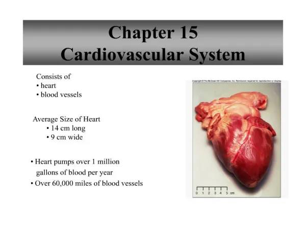
Functional Anatomy • Heart is hollow, four chambered, muscular, roughly fist-sized • Lies just behind the sternum, two thirds lie to left, between the second through the sixth ribs • Heart apex at fifth intercostal space • Surface grooves (sulci) mark the boundaries of the heart chambers
Functional Anatomy (cont.) • Pericardium • Double walled sac enclosing heart • Pericarduim’s structure • Outer fibrous layer: Tough connective tissue, loose-fitting, inelastic sac surrounding heart • Inner serous layer: thinner, more delicate • Serous pericardium: Consisting of two layers: • Parietal layer: Inner lining of fibrous pericardium • Visceral layer (epicardium): covering outer surface of heart & great vessels
The Heart • Pericardial fluid • Thin layer of fluid separating parietal & visceral pericardium • Helps minimize friction during contraction & expansion • Pericardial effusion • Abnormal amount of accumulated fluid between layers
The Heart • Cardiac tamponade • Large pericardial effusion may affect pumping function • Can cause serious drop in blood flow to body • May ultimately lead to shock & death • Pericarditis • Inflammation of pericardium
What is the function of the pericardial fluid ? • helps minimize friction during heart contraction & expansion • provide protection against trauma • mark the boundaries of the chambers of the heart • prevents atrial backflow during ventricular contraction
The Heart (cont.) • Heart wall is composed of 3 layers: • Outer: epicardium • Middle: myocardium comprises bulk of heart & is composed of muscle tissue • Inner: Endocardium • Forms thin continuous tissue with blood vessels • Heart forms 4 muscular chambers • Upper chambers, right & left atria • Lower chambers are right & left ventricles • Responsible for forward movement of blood
Atrioventricular Valves • AV valves lie between atria & ventricles • Tricuspid valve is at right atrium exit • Mitral valve at left atrium exit • Ventricular contraction forces valves closed, preventing backflow of blood into atria • Lower ends of valves anchor to ventricular papillary muscles by chordae tendineae • Papillary contraction during systole pulls on chordae, preventing valve reversing into atria
What is the role of ventricular contraction ? • relaxes chordae tendineae, preventing valves reversing into atria • to rest the heart muscle • to forward movement of blood • prevents atrial blood backflow
Semilunar Valves • Consist of 3 half-moonshaped cusps • Separates ventricles from their arterial outflow tracts, pulmonary artery & Aorta • Situated at ventricle exits to outflow tracks (arterial trunks) • Pulmonary valve lies between right ventricle & pulmonary artery • Aortic valve lies between left ventricle & aorta
Semilunar Valves (cont.) • Systole: (cardiac contraction) valves open, allowing ventricular ejection into arteries (pulmonary artery and aorta) • Diastole: valves close preventing back flow of blood into ventricles
Coronary Circulation • Heart’s high metabolic demands require an extensive circulatory system • The heart requires more blood flow per gram of tissue weight than any other organ besides kidneys
Coronary Circulation (cont.) • Right & left coronary arteries arise under aortic valve cusps • Coronary artery pressure becomes higher than aortic pressure during systole • Prevents flow of blood into coronaries • Diastole is when coronary blood flow occurs thus, diastolic pressure is very important
Left Coronary Artery (LCA) • Positioned underneath aortic semilunar valves • LCA branches into: • Left anterior descending (LAD): courses between left & right ventricles • Circumflex: courses around left side of heart between left atrium & left ventricle
LCA (cont.) • LCA provides blood to left atrium, left ventricle, majority of interventricular septum, half of interatrial septum, & part of right atrium • See Figure 9-4 & Table 9-1
Right Coronary Artery (RCA) • RCA proceeds around right side of heart between right atrium & right ventricle • Many small branches as RCA moves around right ventricle • RCA ends in its posterior descending (RPD) branch, which courses between right & left ventricles. • Provides blood flow to most of right ventricle & right atrium, including sinus node • See Figure 9-4 & Table 9-1
Problems With Coronary Blood Flow • Myocardial Ischemia • Partial obstruction of coronary artery • Decreasing oxygen supply to tissue • a.k.a. Angina Pectoris • Myocardial Infarction (MI) • Sometimes called “infarct” • Complete obstruction of coronary artery • Causes death of heart tissue
Coronary Veins • Collect venous blood after passing through myocardial capillary bed • Veins closely parallel coronary arteries • Great cardiac vein follows LAD • Small cardiac vein follows RCA • Left posterior vein follows circumflex • Middle vein follows RPD • These all come together to form coronary sinus, which empties into right atrium • Thebesian veins drain some coronary venous blood into all heart chambers • Those draining into left atrium & left ventricle bypass lungs, creating an anatomic shunt • Normal anatomic shunt = 2-3% of total cardiac output
Properties of Heart Muscle • Heart’s ability to pump depends on: • Initiating & conducting electrical impulses • Synchronous myocardial contraction • Made possible by key properties of myocardial tissue • Excitability: ability to respond to stimuli • Inherent rhythmicity: initiation of spontaneous electrical impulse • Conductivity: spreads impulses quickly • Contractility: contraction in response to electrical impulse • Unique featurecannot go into tetany
Properties of Heart Muscle (cont.) • Refractory period • Time period myocardium cannot be stimulated • Lasts 250 milliseconds; nearly as long as systole
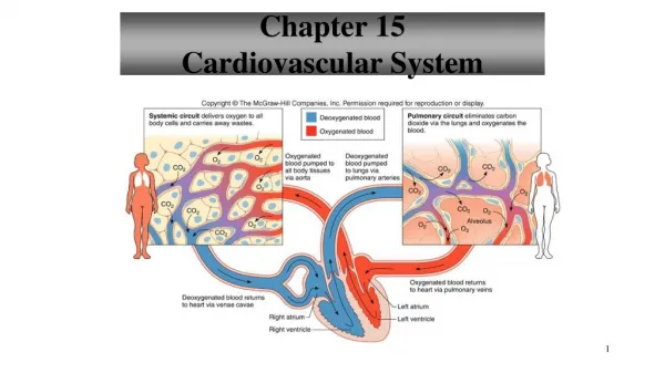
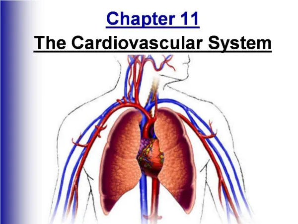
The Vascular System • Composed of 2 major subdivisions: • Systemic vasculature • Pulmonary vasculature • Systemic vasculature begins with aorta on left ventricle & ends in right atrium • Pulmonary vasculature begins with pulmonary trunk out of right ventricle & ends in left atrium • Systemic venous blood returns to right atrium via: • Superior vena cava (SVC): drains upper extremities & head • Inferior vena cava (IVC): drains lower body • Blood flows through tricuspid valve into right ventricle
The Vascular System (cont.) • Pumped from right ventricle through pulmonary valve into pulmonary artery, which carries it to lungs (oxygenation) • Pulmonary arterial blood returns via pulmonary veins to left atrium • From left atrium oxygenated blood flows through mitral valve into left ventricle • Left ventricle pumps the blood out through aortic valve into systemic circulation • Blood passes through systemic capillary beds into systemic veins & back to SVC & IVC
Where does systemic vasculature begin & end? • begins in the right atrium & ends in the right ventricle • begins in the aorta on the left ventricle & ends in the right atrium. • begins in the pulmonary trunk out of the right ventricle & ends in the left atrium. • begins the left atrium & ends in the inferior vena cava
Systemic Vasculature • 3 components: • Arterial system (conductance vessels) • Large elastic low resistance arteries • Small muscular arterioles (resistance vessels) • Major role in distribution & regulation of blood pressure • Like faucets, control local blood flow into capillaries • Capillary system, microcirculation (exchange vessels) • Transfer of nutrients & waste products • Venous system (capacitance vessels) • Reservoir for circulatory system • Generally holds 3/4 of body’s blood volume • Conduct blood back to heart
Vascular Resistance • Sum of all opposing forces to blood flow through systemic circulation is systemic vascular resistance (SVR) • SVR = Change (Δ) in pressure from beginning to end of system, divided by flow SVR = (MAP – RAP)/CO Where: MAP = mean aortic pressure RAP = right atrial pressure or CVP CO = cardiac output
Pulmonary Vascular Resistance (PVR) • PVR: sum of all opposing forces to blood flow through pulmonary circulation • PVR then calculated as is SVR (ΔP/flow) PVR = (MPAP – LAP)/CO Where: MPAP = mean pulmonary artery pressure LAP = left atrial pressure or wedge pressure CO = cardiac output • PVR: normally much lower than SVR as pulmonary system is low pressure, low resistance
Determinants of Blood Pressure (BP) • Normal CV function maintains blood flow throughout body • Under changing conditions, need constant BP MAP = CO × SVR And MAP = Volume/Capacity • To maintain BP, capacity must vary inversely with CO or volume
Determinants BP(cont.) • Normal adult: MAP values range: • 80 to 100 mm Hg • If MAP falls significantly below 60 mm Hg • Perfusion to brain & kidneys is severely compromised • Organ failure may occur in minutes
Control of Cardiovascular System • The heart works as a demand pump • CV system may alter capacity - how much blood it holds • Decreased capacity results in greater venous return & greater CO • CV system tells heart how much to pump • Accomplished by local & central control mechanisms • Heart plays secondary role in regulating blood flow • Blood flow through large veins can also be affected by abdominal & intrathoracic pressure changes
Cardiac Output (CO) & its Regulation • CO = total amount of blood pumped by heart per minute • CO = Heart rate (HR) × stroke volume (SV) • Normal CO = 5 L/min • End Diastolic Volume (EDV): blood left in atria
CO & its Regulation (cont.) • End Systolic Volume (ESV): blood left in ventricles • HR is primarily determined by CNS • CO is directly related to HR • HR > 160180 is exception; too little time for filling results in decreased EDV, EF, SV, &, thus, CO • SV is determined by • Preload • Afterload • Contractility • SV = EDV-ESV
Stroke Volume & Preload • Preload essentially equals venous return • Amount of volume & pressure at end diastole (EDV, EDP) stretches myocardium • Greater the stretch, the stronger the contraction • Frank-Starling Law • Normal EDV is ~110120 ml • Normal SV is ~70 ml • Ejection fraction (EF) = SV/EDV • Normal ~65% • If it falls to 30% range or below, patient’s exercise tolerance becomes severely limited
Stroke Volume & Preload (cont.) • Afterload: resistance against which ventricles pump, so more afterload makes it harder for ventricles to eject SV • RV afterload = PVR • LV afterload = SVR • All else constant, increase in vascular resistance would decrease SV • Usually does not occur as contractility increases to maintain SV & thus CO
Stroke Volume & Preload (cont.) • Afterload represents sum of all external factors opposing ventricular ejection • Tension in ventricular wall • Peripheral resistance or impedance
Stroke Volume & Contractility • Contractility: amount of force myocardium produces at any EDV • Increased contractility results in greater EF for any EDV • Called positive inotropism • If afterload & contractility increase together, SV is maintained • Positive inotropes • Drugs that increase contractility of heart muscle • Negative inotropes • Drugs decreasing contractility of heart muscle
If a patient is given Dobutamine, a positive inotropic drug, how is contraction of the heart affected? • decrease the force of contractions • increase the force of contractions • not affect the force of contractions • intermittently increase & decrease the force of contractions
Cardiovascular Control Mechanisms • Integration of local & central mechanisms to ensure all tissues have enough blood flow • Normally, local control is primary determinant • With large changes in demand, central control becomes primary • Central control in medulla has areas for: • Vasoconstrictionincreases adrenergic output • Vasodepressorinhibits vasoconstrictor center • Cardioacceleratoryincreases heart rate • Cardioinhibitorydecrease heart rate (by increasing vagal stimulation to heart)