Uropoetic system
Uropoetic system. Histology and embryology. Uropoetic system. Kidney ( Ren ) Nephrone ( Nephron ) Collecting tubules ( Tubuli colligentes ) Callices ( Calices renales ) Renal pelvis ( Pelvis renalis ) Ureter ( Ureter ) Urinary bladder ( Vesica urinaria ) Ur ethra ( Urethra ).


Uropoetic system
E N D
Presentation Transcript
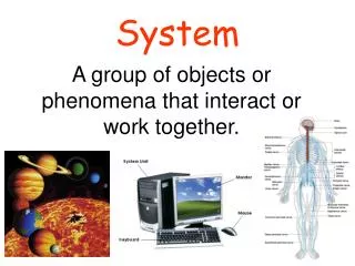
Uropoetic system Histology and embryology
Uropoetic system • Kidney (Ren) • Nephrone (Nephron) • Collecting tubules (Tubuli colligentes) • Callices (Calices renales) • Renal pelvis (Pelvis renalis) • Ureter (Ureter) • Urinary bladder (Vesica urinaria) • Urethra (Urethra)
Development of urinary system • Development of kidney • Pronephros • Mesonephros • Metanephros • Development of excretory passages
Pronephros • Intermedial mesoderm of cranial 12-13 somites • From 21st day (4 somites) • Rudimental, soon disappear • Persistent ductus Wolffi
Mesonephros • From intermedial mesoderm • Sac elongates to ductus Wolffi • In stage of 27-28 somites ingrowths to cloaca • From about 23rd day • With glomerulum and collective tubule • Caudal part is origin for gonads
Metanephros • Definitive stage of kidney • nephrogenic blastema 3rd to 5th lumb. somites • Development begins at the end of 5th week • Relative ascent during development • Ureteral bud grows to blastema • Ureteral bud – ureter, pelvis, large and small calices and collecting tubules • metanephrogenic blastema - nephrone
Induction of nephrons • Interaction between blastema and ureteric bud • Connecting with the branch of ureteral bud • Later development of nephrone • Distance of glomerulum from the renal medulla is the function of age • Oldest nephrons – juxtamedular
Development of excretory passages • Urinary bladder • Developed from cranial part of urogenital sinus • Urachus • Separation of excretory passages for urine and genital ducts • Urethra • From medial part of urogenital sinus (at females) • From medial and caudal part (at males) and glandular plate
Kidney (Ren) • Nephron • Intersticium • Juxtaglomerular apparatus • Blood supply • Intrarenal excretory passages for urine
Nepron • Renal body • Glomerulum • Bowman‘s capsule • Proximal tubule • Intermedial tubule • Distal tubule
Bowman‘s capsule Parietal part (squamous epitelium) Visceral part (podocytes - pedicles) Glomerulum a. afferens a. efferens fenestrated capilaries w/o diaphragm Renal body (Malpighi) • Filtration barier • basal membrane - collagen type IV and heparan sulfate • Mesangial cells
Proximal tubule • Single layered cubic epitelium • Brush border on apical part • Basolateral labyrinth • Abundand mitochondrias • Resorption of NaCl and water (80-95%) • Na+ into cell pasive, out active
Intermedial tubule • Thin limb of Henle‘s loop • Squamous cells, poorly organels • Descendent part permebile for water • Ascendent part inpermeabile for water — • Juxtamedular nephrons have long HL • countercurrent multiplier - hyperosmotic medulla
Distal tubule • Cells are lower than in proximal tubule • No brush border • Basolateral labyrinth • Reabsorption of natrium, secretion of kalium • macula densa - chemoreceptors (Cl-)
Juxtaglomerular apparatus • Parts: • macula densa of distal tubule • granular cells of afferens arteriol (also efferens) • mesangial cells • Function: • Regulation of blood pressure - renin
Intersticium • Connected with basal lamina of vessels and tubules • Cortical vs. medullar • Cells: • Fibroblast-like cells • Cells with lipid droplets • Macrophages, pericytes • Non-cellular elements • proteoglycans, glycoproteins, interstitial fluid
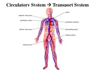
aa. interlobares aa. arcuatae aa. corticales radiatae aa. capsulares aa. afferentes aa. efferentes Peritubular capillaries vasa recta vv. arcuatae vv. corticales radiatae vv. stellatae vv. interlobares Blood supply
Intrarenal excretory passages • Collecting tubules (Tubulli colligentes) • Single layered cubic epithelium • Round nucleus, light cytoplasm • Papilar ducts (Ductus pappilares) • Single layered columnar epithelium • Light cytoplasm • Regulated water reabsorption (ADH)
Slides Slide U1- kidney HE Slide U2 - kidney Van Gieson
Diabetic nephropathy Acute pyelonephritis Renal diseases
Excretory passages • Calices and pelvis • Ureters • Urinary bladder • Urethra
Excretory passages • Mucosa (tunica mucosa) • epithelium transitional • lamina propria mucosae (collagen) • Smooth muscle (tunica muscularis) • Adventitia (tunica adventitia)
Renal pelvis and calices • 2-3 calices in every pelvis • 2-3-layered transitional epithelium • Smooth muscle cells spirally
Ureter • Folded mucosa • Smooth muscle • Inner layer longitudinal • Outer layer circular
Slides Slide U3 - ureter HE
Urinary bladder • Mucosa folded except of area trigonum vesicae • 3-layered smooth muscle layer • inner plexiform • middle circular ( m. sphincter vesicae) • outer longitudinal • Part of bladder covered by serosa (peritoneum)
Slides Slide U4 – urinary bladder HE Slide U5 – urinary bladder Van Gieson
Urethra • Male urethra have 4 parts: • First two part covered by transitional epithelium • Other two covered by multilayered sqaumous epithelium • Female urethra • Transitional epithelium in intramural part, continuing with multilayered squamous