Download
1 / 41
410 likes | 428 Views
This resource module provides information on the significance of colon cancer in the elderly, factors and conditions associated with the disease, guidelines for screening and surveillance, and various treatment options for colorectal cancer. Learn about the prevalence and risk factors, clinical presentation, diagnostic tests, tumor markers, and TNM staging classification.

E N D
Learning Objectives • Describe the significance of colon cancer in the elderly • Identify the factors and conditions associated with the colon cancer • Describe the appropriate guidelines for screening and surveillance for the early detection of cancer • Describe the various treatment options
Colorectal Cancer • 90% of cases occurs after age 50. • Third leading cause of cancer in the US • Second leading cause of cancer death • Average lifetime risk for developing this cancer is 6% • Men and women are affected equally • Women are more likely to have right sided colonic adenomas • Distributed evenly among various racial groups • African Americans and Hispanics have lower survival rate
Risk Factors • Age >50 yrs • High fat, low fiber diet • IBS – Chronic ulcerative colitis and Crohn’s disease • Familial adenomatous polyposis (FAP) • Heriditary nonpolyposis colorecal cancer (HNPCC) • Hamartomatous polyposis syndromes • Peutz-Jeghers syndrome • Juvenile polyposis • Family history – Colorectal adenomas, Colorectal cancer • Personal history of Colorectal adenomas, Ureterosigmoidostomy, Breast, Ovarian and Uterine cancers
Familial Risk Approximate Familial setting Life time risk of Colon Ca Gen population risk in US 6% 1 first degree relative with colon ca Two-Three fold increase 2 first degree relatives with colon ca Three-Four fold increase 1st° relative Δ with colon ca ≤ 50 yrs Three-Four fold increase One 2nd or 3rd° relative with colon ca 1.5 fold increase Two 2nd ° relatives with colon ca Two-Three fold increase One 1st ° relative with polyp Two fold increased
Factors associated with increased risk for CRC Lack of physical activity Consumption of red meat Obesity Cigarette smoking Alcohol use Factors associated with decreased risk for CRC MVI containing folic acid ASA and other NSAID’s Post menopausal HRT Ca supplementation Selenium Consumption of fruits, vegetables and fiber
Clinical Presentation • Depends on tumor location • Proximal (right sided) lesions present with symptoms caused by anemia – fatigue, weight loss, shortness of breath, lightheadedness, mahagony feces caused by occult bleeding • Distal (left sided) lesions present with symptoms of obstruction, changes in BM pattern, postprandial colicky abdominal pain, hematochezia
CLINICAL MANIFESTATIONS • Abdominal pain 44% • Change in bowel habit 43% • Hematochezia or melena 40% • Weakness 20% • Anemia without other gastrointestinal symptoms 11% • Weight loss 6% • Some patients have more than one abnormality • 15 to 20% of patients have distant metastatic disease at the time of presentation
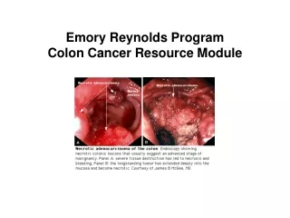
Diagnostic Tests • Digital rectal exam (DRE) • Barium enema (BE) with or without air contrast: used primarily to locate deformities of intestinal topography • Sigmoidoscopy, rigid type or flexible fiber optic type: used to visualize local rectal tumors or for routine screening • Colonoscopy (or colon endoscopy): Direct visual examination of the colon and rectum detects early polypoid tumors preoperatively and recurrences post-resection; Multiple biopsies may be performed at time of study to increase sensitivity • Computed tomography (CT): Used to stage disease and identify metastases • Transrectal ultrasound (TRUS): An excellent choice for preoperative staging of rectal carcinomas • Magnetic resonance imaging (MRI): very useful for diagnosing metastatic disease • Laparotomy: Useful in detecting metastases to abdominal regions (especially omentum or liver) that often remain undetected by current imaging techniques
Barium enema (BE) with or without air contrast: used primarily to locate deformities of intestinal topography
Tumor markers Carcinoembryonic antigen (CEA) Carbohydrate antigen (CA) 19 9, CA 50, and CA 195 • Have a low diagnostic ability to detect primary CRC overlap with benign disease • low sensitivity for early stage disease • An expert panel on tumor markers convened by the American Society of Clinical Oncology (ASCO) recommended that serum CEA levels not be used as a screening test for colorectal cancer • Have prognostic utility
TNM Staging Classification of CRC Primary tumor (T) • TX - Primary tumor cannot be assessed • T0 - No evidence of primary tumor • Tis - Carcinoma in situ: intraepithelial or invasion of lamina propria • T1 - Tumor invades submucosa • T2 - Tumor invades muscularis propria • T3 - Tumor invades through the muscularis propria into the subserosa or into nonperitonealized pericolic or perirectal tissues • T4 - Tumor directly invades other organs or structures, and/or perforates visceral peritoneum • Regional lymph nodes (N) • NX - Regional lymph nodes cannot be assessed • N0 - No regional lymph-node metastasis • N1 - Metastasis in 1 to 3 regional lymph nodes • N2 - Metastasis in 4 or more regional lymph nodes • Distant metastasis (M) • MX - Distant metastasis cannot be assessed • M0 - No distant metastasis • M1 - Distant metastasis
Prognosis and 5 yr survival rates for Colon Cancer • Stage I (T1-2N0) - 93% • Stage IIA (T3N0) - 85% • Stage IIB (T4N0) - 72% • Stage IIIA (T1-2 N1) - 83% • Stage IIIB (T3-4 N1) - 64% • Stage IIIC (N2) - 44% • Stage IV - 8%
Guidelines for screening average risk adults aged 50 years or older • Fecal occult blood test (FOBT) every year • Occult stool testing must be repeated at least 3 times on different stool samples. • Diet must be free of peroxidase activity (turnips & horseradish). • Tests may need to be repeated if there is a history of: • Usage of possible gastric irritants such as salicylates, other anti-inflammatory agents • Hemorrhoids • Diverticulitis • Peptic ulcer disease (PUD) or other cause of GI bleeding
Guidelines for screening average risk adults aged 50 years or older • Fecal occult blood test (FOBT) every year • Flexible sigmoidoscopy every five years • FOBT every year combined with flexible sigmoidoscopy every five years. • Double-contrast barium enema every five years • Colonoscopy every ten years
Key elements in screening average risk people • Symptoms require diagnostic work up • Offer screening to men and women aged 50 and older • Stratify patients by risk • Options should be offered • Follow up of positive screening test with diagnostic colonoscopy • Appropriate and timely surgery for detected cancers • Follow up surveillance required after polypectomy and surgery • Providers need to be proficient • Encourage participation of patients
Screening for high-risk people • A first-degree relative (sibling, parent, child) who has had colorectal cancer or an adenomatous polyp: Screening should begin at age 40 years • Family history of familial adenomatous polyposis (FAP): Screening should begin at puberty Sigmoidoscopy - annually, beginning at age 10 to 12 years Colonoscopy - every five years • Family history of hereditary nonpolyposis colorectal cancer (HNPCC): Screening should begin at age 21 years Sigmoidoscopy - annually, beginning at age 10 to 12 years Colonoscopy - every one to two years, beginning at age 20 to 25 years or 10 years younger than the earliest case in the family, whichever comes first • Personal history of adenomatous polyps Screening should be based on pathological findings Advanced or multiple adenomas (3 or greater): First follow-up colonoscopy should occur in 3 yrs 1 or 2 small (< 1 cm) tubular adenomas: First follow-up colonoscopy should occur at 5 years • Personal history of colorectal cancer: After colon resection Approximately six months after the surgery If the colonoscopy performed at six months is normal, subsequent colonoscopy should be repeated at 3 years and then if normal, every 5 years • Personal history of inflammatory bowel disease Every one to two years after an eight year history of the disease with pancolitis or Every one to two years after 15 years history of left-sided colitis or For all patients beginning with eight to ten years of disease to document the extent of the disease
Blood work that may be indicated • Complete blood count (CBC) • Liver chemistries: Abnormal liver enzyme results may suggest metastatic disease • Carcinoembryonic antigen level (CEA) - Normal value: 0-2.5 mg/ml; up to 10 mg/ml in tobacco smokers Useful in establishing diagnosis and recurrence for tumors that secrete CEA and in following disease progression. Because colon lesions are not likely to secrete CEA, it is not a highly reliable indicator of colon cancer. If CEA is elevated, return to normal levels is expected to occur within 48 hours after complete tumor excision • C-Reactive protein (CRP) Increased plasma concentrations of CRP is associated with subsequent development of colon cancer Preliminary findings are consistent with the established association between colon cancer and inflammatory bowel disease (IBD) • CRP research is ongoing and full corroboration of suggestive findings has not been established
Genetic testing • Genotyping (APC gene test) should be used when other diagnostic avenues are exhausted • Medically necessary in presence of strong family history for familial adenomatous polyposis (FAP), attenuated familial adenomatous polyposis (AFAP), or hereditary nonpolyposis colorectal cancer (HNPCC)
Malignant lesions Adenocarcinoma Lymphoma Carcinoid tumor Kaposi’s sarcoma Prostate cancer Benign lesions Crohn’s colitis Diverticulosis Endometriosis Solitary rectal ulcer Lipoma Tuberculosis Amebiasis Cytomegalovirus Fungal infection Extrensic lesion Arterio-venous malformations Adenomatous polyps- Premalignant neoplasm Morphological types- tubular, tubulovillous, villous Ischemic colitis Infarcted colon Megacolon Hemorrhoids Differential diagnosis
Treatment Options Surgical excision: Mainstay of curative Rx • Specific procedure depends on the anatomic location of the cancer, but typically involves hemicolectomy • Surgical resection of affected bowel with clear margins, along with the adjacent mesentery and at least 12 regional nodes • For rectal tumors, total mesorectal excision with a distal surgical margin of at least 2 cm is recommended • For tumors that are located within 6 cm of the anal verge, or involve the anal sphincter, wide surgical resection with abdomino-perineal resection and permanent colostomy is recommended • Local excision, for palliative treatment or simple polyp removal Radiation therapy: • Postoperative radiation, with or without chemotherapy, significantly reduces local recurrence rates • Common regimen incorporates infusional 5-fluorouracil (5-FU) as a radiosensitizer to boost the efficacy of pelvic radiation • Administered as 45 to 55 Gy over 5 weeks • Repeated as needed
Treatment Options SYSTEMIC CHEMOTHERAPY • 5-FU has been the mainstay of systemic chemotherapy for CRC • Capecitabine was approved in 2001 as first-line therapy for metastatic CRC • Irinotecan (Camptosar), Oxaliplatin (Eloxatin), Bevacizumab, Cetuximab Electrocoagulation • Mostly palliative treatment for rectal carcinomas • Curative for small subset of patients
History and physical examination: Every 3 to 6 months for the first 3 yrs Every 6 months during years 4 and 5 Then anually thereafter CEA: Every 3 months for at least 3 yrs in pts with stage II or III CRC if they are candidates for surgery or systemic therapy LFT’s, CBC, CXR and Fecal occult blood test: Not recommended CT of chest and abdomen: Pts with CRC at higher risk for recurrence (stage III or II with multiple poor risk features) should undergo annual CT of chest and abdomen for 3 yrs if they are eligible for curative intent surgery Pelvic Imaging: Annual pelvic CT should be considered for rectal surveillance, particularly if the pt has not been treated with pelvic radiation therapy Colonoscopy: In the pre-operative or post-operative setting to document a cancer free or polyp free colon Pts presenting with an obstructive cancer should undergo colonoscopy within 6 months of surgery. Repeat colonoscopy is recommended at 3 yrs, and if normal every 5 yrs thereafter Flexible Proctosigmoidoscopy: Every 6 months for 5 yrs in pts who have not received pelvic radiation therapy, Summary of Updated 2005 CRC Surveillance Guidelines from the American Society of Clinical Oncology
Bibliography • Colon cancer screening, surveillance, prevention, and treatment: conventional and novel technologies Cappell MS - Med Clin North Am - 2005 Jan; • The pathophysiology, clinical presentation, and diagnosis of colon cancer and adenomatous polyps.Cappell MS - Med Clin North Am - 01-JAN-2005; 89(1): 1-42, viiFrom NIH/NLM MEDLINEScreening of patients at average risk for colon cancer.Mandel JS - Med Clin North Am - 01-JAN-2005; 89(1): 43-59, vii • Surveillance of patients at high risk for colorectal cancer.Syngal S - Med Clin North Am - 01-JAN-2005; 89(1): 61-84, vii-viii • Prevention and therapy of colorectal cancer.Hawk ET - Med Clin North Am - 01-JAN-2005; 89(1): 85-110, viii • http://www.rand.org/pubs/monograph_reports/MR1281/index.html • http://www.muschealth.com/cancer/tools/hassessment.htm#colon • www.rand.org/pubs/working_papers/2006/RAND_WR174-1.pdf • www.rand.org/pubs/working_papers/2005/RAND_WR178.pdf • http://www.gastrojournal.org/article/PIIS0016508502158951/fulltext • http://guidelines.gov/summary/summary.aspx?doc_id=4006&nbr=003135&string=colon+AND+cancer+AND+screening
Patient education web sites and resources • National cancer institute 1-800-4-cancer, www.nci.nih.gov • The American Society of Clinical Oncology, www.asco.org • National comprehensive cancer network, www.nccn.org/patients_gls.asp • American cancer society, 1-800-acs-2345, www.cancer.org • National library of medicine, www.nlm.nih.gov/medlineplus • The american gastroenterological association, www.gastro.org • The american college of gastroenterology, www.acg.gi.org