Download
1 / 41
440 likes | 826 Views
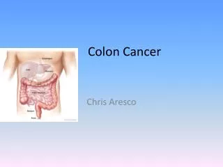
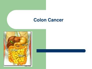
Colon Cancer. Epidemiology. 3 rd most common cancer in males and females. Accounts for 11% of cancer deaths. In 2000, 130,200 cases (colon and rectum). Lifetime risk 6%. Epidemiology. Rare before the age of 40y, rapid increase at 50y.

E N D
Epidemiology • 3rd most common cancer in males and females. • Accounts for 11% of cancer deaths. • In 2000, 130,200 cases (colon and rectum). • Lifetime risk 6%.
Epidemiology • Rare before the age of 40y, rapid increase at 50y. • At presentation 37% localized, 37% regional, 20% metastatic. • 1 and 5y survival is 80% and 61% overall. • IBD, FAP, HNPCC, FHX are at inc risk • 75% sporadic, rest are in those at high risk.
Risk Factors • Diet- red meat, animal fat, increased cholesterol in stool. Folate is protective. Decreased folate= Kras mutations. • Calcium supplementation decrease new adenomas. • Fiber- use not supported yet for protection from cancer. • Meds-HRT, ASA, NSAIDS, COX2 protective and in some cases cause regression of polyps. • Alcohol consumption increases risk.
Risk Factors • Polyps-Most cancers arise from them. • Classified as neoplastic (adenomatous)which are benign or malignant, and nonneoplastic (hyperplastic, mucosal, inflammatory, hamartomaous). • Adenomatous polyps found in 33% of people by age 50, 50% by age 70. • Most lesions <1cm, 60% single, 40% multiple. • Invasive cancer will develop in 24% when untreated.
Polyps • Three variants: Tubular(75-87%), tubulovillous (8-15%), Villous(5-10%). • Tubulovillous, villous(most in rectum) have most increased risk of cancer 20% and 40% respectively. • Size, degree of dysplasia (46% cancer >2cm, 34% in severe dysplasia).
Polyps • Poor differentiation, lymphovascular invasion, submucosal, positive margin. • Level of invasion 0-4 head to stalk (Haggitt level) • Lymphatic channels do not penetrate above the muscularis mucosa, so level 4 most important. • 8-17% of polyps with invasive carcinoma will have + nodes. • A negative margin of resection associated with decreased adverse outcome (0.8%). 27% of patients with positive margins will have adverse outcomes.
Treatment • Endoscopic removal, surveillance every three years. • Biopsy if it can’t be removed. • Surgery for those not amenable to safe polypectomy (large sessile villous lesions).
Treatment • Fungation, ulceration, distortion are contraindications for polypectomy. • Colectomy indicated for residual carcinoma, those at high risk for +LN despite complete polypectomy. • +margin, poor diff, level 4, vascular, lymphatic invasion. • Sessile polyp with invasive cancer should be considered for resection even if no high risk pathologic features. • Weigh all against pts medical condition of course.
Hereditary Polyposis Syndromes • All have this in common: Multiple intestinal polyps, extraintestinal manifestations. • FAP: 1-2% of colon cancer patients. A point mutation of APC gene on chromosome 5, band q21. • Polyps found throughout the GI tract but most in colon. Symptoms manifest by ages 16-50. • Cancer will develop in all by age 50.
Hereditary Polyposis Syndromes • Gardner’s Syndrome: Variant of FAP. Colonic and extracolonic manifestations. • Periampulary lesions, duodenal lesions, gastric polyps. • Ocular, cutaneous, skeletal (retinal, mandible, jaw, teeth, sebaceous cysts). • Desmoids, hepatoblastoma, thyroid cancer, Turcot’s syndrome (brain).
Hereditary Nonpolyposis Syndromes • Lynch I and II. Occurs five times more frequently than familial polyposis. 1-5 % of colon cancers. Lynch I just colon, Lynch II also involves endometrium, ovary, stomach, small bowel, biliary, pancreas, ureter, renal pelvis. • 85% lifetime risk of colon cancer, more right sided cancers (60-70%), earlier (45y), lower stage, better survival, but 20% risk of metachronous, synchronous lesions.
Inflammatory Bowel Disease • Ulcerative colitis carries a risk of colorectal carcinoma 30 times greater than general population. • Risk increases with duration of disease. • After 30 years, risk increases to 35% • Crohn’s disease associated with 10-20 fold increased risk of cancer. • Need to do surveillance in these population.
Previous Colon Cancer • A second primary colon cancer is three times more likely to develop in patients with a history of colon cancer. • Metachronous lesions develop in 5-8% of patients.
History of First-Degree Relatives • People with first-degree relatives with colorectal cancer have a 1.8-8 fold increase risk of colorectal cancer. • Risk is higher if more than one relative affected. • Risk is higher if developed in the relative at a young age.
Screening • FOBT, DCBE, endoscopy most useful screening methods. • FOBT detects cancer at an earlier stage, with reduction in cancer deaths. • Flexible sigmoidoscopy and polyp clearance has resulted in decreased colon cancer. • Value of full colonoscopy is noted since 40% of colon cancers occur proximal to splenic flexure. • DCBE used if pt refuses scope, or poor scope, etc.
Screening • CEA has no role in in screening for primary lesions. False positives occur in benign disease(lung, liver, bowel) as well as malignancies of pancreas, breast ovaries, prostate, head and neck, bladder, kidney. • CEA increased in smokers. • 60% of tumors will be missed by CEA alone.
Recommendations • Age>50 asymptomatic, average risk. • FOBT yearly, scope if positive • Flex sigmoidoscopy every 5y (full colon if +) • Increased risk: Same but start age 40.
Recommendations • Hx of HNPCC: Full colon every 1-2y (20-30y) then full colon yearly after 40y. • Hx Aden Polyps: repeat in 3y, second exam normal repeat 5y. • Hx Colon cancer: Full colon within 1y, if second normal repeat 3y, if next normal every 5y. • FAP: Counseling, Flex Sigmoid every 12 months.
Pathology • >90% adenocarcinomas. Four morphologic variants. • Ulcerative (most common), exophytic (polypoid, fungating), annular (classic applecore), submucosal infiltrative(linnitus type). • Grading system 1-3. Most developed to least differentiated glandular structures.
Staging- Aston Collier • A- to submucosa only • B1- to muscularis only • B2- thru wall, not adjacent. • B3- Adjacent organs involved. • C1- B1 plus LN • C2- B2 plus LN • C3- B3 plus LN • D- Distant mets
Staging-TNM • T1 invades submucosa • T2 invades muscularis • T3 invades subserosa • T4 invades organs outside • N1- 1-3 nodes • N2- 4 or more nodes • N3- central nodes • M0- no mets • M1- distant mets
Clinical Presentation • Bleeding, pain, bowel habit changes, weight loss, anorexia, nausea, vomiting, fatigue, anemia. • Right upper quadrant pain, fevers sweats, hepatomegaly, ascites, effusions, adenopathy(METS). • Obstruction(5-15%) increases risk of death 1.4 fold. • Perforation (6-8%) increases it 3.4 fold. • Stage I 15%, Stage II 30%, Stage III 20%, Stage IV 25%. • Obstruction less common on right side.
Diagnosis • Scope, CXR, CBC, CEA, U/A, LFTs. • Preop CT scan? Some get it for abnormal LFTs only (but only 15% of liver mets have abnormal LFTs). Others will get it if large bulky tumors to see about adjacent organs, LN. • 10% of mets are missed with preoperative and operative evaluations, IOUS best for this.
Diagnosis • 15-20% liver mets not palpable. • Preop CEA reflects prognosis, disease extent (over 10-20 poor) • CEA may not be elevated in poorly differentiated or rectal cancers. • CEA really only good for follow up.
Rectal Cancer • In addition to H&P, CXR, CBC, LFTs, U/A, EUS, Proctoscopic exam, full colonoscopy, CT scan should be done for rectal cancer. • Accurate preoperative staging critical because stage may influence treatment decisions such as trans anal excision, preop chemoradiation.
Rectal Cancer • EUS is most accurate tool in determining tumor stage with all layers identified with 67-93% accuracy. • Differentiating T1 from T3 easy but T2 from T3 harder. • Limitations of EUS: operator experience, differentiating LN vs.blood vessels, post radiation changes, stenotic lesions, overstaging (10-15%), understaging (1-2%). • Superior to CT or MRI for depth of tumor.
Rectal Cancer • Lymph node staging more difficult. EUS 62-83% accurate, CT scan 35-73% accurate. • All these tests pick up size of LN only. • 50-75% of involved LN are normal in size, so may not be picked up. Similarly, enlarged LN may be inflammatory, so false negative. • LN> 3mm and hypoechoic are likely to have malignancy, also FNA might help under EUS guidance.
Rectal Cancer • CT scanning of abdomen and pelvis is important for other organ involvement, and distant spread. • CT is better than EUS for contiguous organ involvement.