Download
1 / 122
1.22k likes | 1.24k Views
Learn about the important functions of bones in the body, including support, protection, leverage, storage, and blood cell formation. Explore the two main types of bone, the structure of bone cells, the blood supply to bone, and bone formation. Discover the different shapes of bones and the role of bone marrow. Gain knowledge on common bone features such as articular surfaces.

E N D
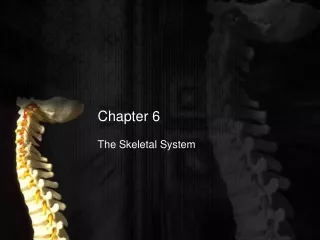
Chapter 6 The Skeletal System
Functions of Bone • Support and Protection • It provides a framework for the body and gives it shape. • It supports and protects organs from injury. • Leverage • It provides a place for muscles, tendons, ligaments and tendons of the body to attach. • It helps make movement possible. • Storage • It stores minerals (Calcium) • Blood Cell Formation • It provides a place for hemotpoeisis
Bone Structure • Two main types of bone: • Cancellous Bone • Light and spongy inner layer of bone. • Consists of tiny spicules with bone marrow between. • Provides strength but prevents damage. • Compact Bone • Heavy and dense layer of outer bone • Found in shafts of long bones • Composed of haversian systems around a haversian canal
Bone Structure continued • Osteocytes- bone cells. • Periosteum- covering of bone • Inner layer contains osteoblasts. • Endosteum- membrane that lines the hollow interior surface of bones.
Haversian Systems Concentric layers of ossified bone matrix arranged around a central Haversian canal Layers of ossified bone matrix
Bone Cells • Osteoblasts • Cells that form bone. • Osteocytes • Once osteoblasts are trapped inside matrix of osseous material. • Can revert back to osteoblasts (remember this from last chapter?) • Osteoclasts • Eat bone away. • Are the remodelers of bone • Withdraw calcium when needed from bone • Why is this important?
Blood Supply to Bone • Tiny vessels penetrate the periosteum. • Volkmann’s canals- tiny channels in the bone matrix that vessels pass through. • Are at right angles to Haversian canals that run lengthwise in the bone. • Nutrient Foramina- Where large vessels enter the bone. • Carry blood into and out of bone marrow. • Can be mistaken for fracture on radiographs.
Bone Formation • Bone is formed in 2 ways: • Endochondral Bone formation • Intramembranous Bone formation • Bone formation and growth is stimulated by Growth Hormone (GH) from the anterior pituitary (adenohypophysis) gland in the brain.
Endochondral Bone Formation • Endochondral Bone formation • Cartilage bone formation • Cartilage first, then bone • How most bones develop • Start as cartilage rods in long bones in diaphysis (shaft). • Contains primary growth center • Cartilage is removed gradually as bone is created and growth center expands. • Secondary growth center • Develop in epiphysis (ends) of bones
Growth plates • Located between diaphysis and epiphysis. • May be called epiphyseal growth plates. • Sites of creation of new bone that allows bone to lengthen as animal grows. • Cartilage is created on epiphyseal side while bone is created on diaphyseal side. • When bone reaches full length, all cartilage is replaced by bone and plates “Close”. • Remodeling may take place but bone will not get any longer. • Young animals may have epiphyseal fractures because this area is weaker than rest of bone.
Panosteitis • “Growing Pains” in dogs. • Inflammation of various bone layers • Seen prevalently in young giant breed dogs. • Basically, body can not keep up as bones are growing very quickly. • Can be confirmed through Radiographs. • May cause “Shifting Leg Lameness” • Treated with rest and anti-inflammatories • Usually occurs in the center of the bone.
Intramembranous Bone Formation • Occurs only in certain skull bones • Bone forms in the fibrous membranes that cover the brain in fetus. • Bone forms directly from osteoblasts with no cartilage intermediary.
Bone Shapes • Long Bones • Short Bones • Flat Bones • Irregular Bones
Long Bones • Longer than they are wide. • Has a proximal and distal epiphysis consisting of cancellous bone. • Main part of bone is diaphysis which composed of compact bone. • Found in digits and limbs.
Short Bones • Shaped like cubes. • Have core of cancellous bone covered by compact bone. • Carpal and tarsal bones.
Flat Bones • Thin and flat bones • Consists of two layers of compact bone separated by cancellous bone. • Bones in skull, pelvis, and scapula are examples.
Irregular Bones • Miscellaneous bones that do not fit into another category. • May have characteristics of more than one category. • Include vertebrae and sesamoid bones. • Patella is largest sesamoid bone in body.
Bone Marrow • Fills the spaces within bones • Has two types: • Red bone marrow • Hematopoietic tissue forms new blood cells. • Majority of bone marrow in young animals but less of older animals • Yellow bone marrow • Consists primarily of adipose connective tissue. • Common type of marrow in adult animals • Does not produce blood cells but can revert to red marrow if needed.
Common Bone Features • Articular Surfaces • Joint surfaces where bones come in contact with each other to form joints. • Consists of: • Condyles • Head • Facet • Covered by articular cartilage • Composed of what type of cartilage?
Condyle • Large, round articular surface. • Major condyle is located on end of humerus and femur. • Also located in skull.
Head • Somewhat spherical articular surface on the proximal end of a long bone. • Found on humerus, femur and rib. • Head is usually joined with rest of bone by a neck.
Femoral Head Osteotomy (FHO) • Head of femur is removed in cases of trauma or severe arthritis. • A “false joint” forms which gives more comfort to the patient.
Facet • A flat articular surface. • Found in carpal and tarsal bones as well as in vertebrae, radius and ulna.
Processes • All projections of a bone. • Heads and condyles are considered to be processes. • Tendons may attach to processes
Holes and Depressed Areas • Foramen: A hole in bone. • Usually allow the passage of nerve or blood vessel. • May exist simply to lighten structure (pelvis-obturator foramen) • Fossa: A depressed of sunken area on the surface of a bone. • Usually occupied by muscles or tendons.
Types of Skeletons • Bones of head and trunk are Axial Skeleton • Bones of limbs and appendages are Appendicular Skeleton. • Some animals may have Visceral Skeleton- bones formed in the viscera or soft organs.
Axial Skeletonbones of head & trunk Skull Hyoid bone Spinal column Ribs Sternum
Skull Usually consists of 37 or 38 separate bones Most skull bones joined by sutures (fibrous joint) Mandible is connected to skull by a synovial joint (TMJ)
Skull External bones: • Frontal bones (2) • Occipital bones (1) • Parietal bones (2) • Temporal bones (2) • Incisive (2 ) • Nasal (2) • Maxillary (2) • Zygomatic (2) • Mandible (2) • Palatine (2) • Turbinates (2)
Skull Bones Continued • Categorized by: • Bones of Cranium • Bones of the ear • Bones of the face
Bones of the Cranium • Cranium-portion of skull that surrounds the brain. • External Bones of Cranium: • Frontal Bones (2) • Interparietal Bones (2) • Occipital Bone (1) • Parietal Bones (2) • Temporal Bones (2) • Internal Bones of Cranium: • Ethmoid Bone (1) • Sphenoid Bone (1)
Occipital Bone • Forms caudoventral portion or base of skull, most caudal skull bone. • Important because: • Where spinal cord exits skull • Skull bone that articulates with first cervical (neck) vertebrae. • Foramen Magnum is in center of occipital bone. • Occipital Condyles are on either side of foramen magnum
Interparietal Bones • Small bones located on dorsal midline between occipital and parietal bones • Clearly visible in young animals, may fuse together in older animals.
Parietal Bones • Form the lateral walls of the cranium • Well developed in dogs, cats and humans, but relatively small in horses and cattle.
Temporal Bones • Located ventral to the Parietal bones • Form walls of the cranium • Contain middle and inner ear structures • Form Temporamandibular Joints (TMJ’s) with the mandible (Lower jaw)
Frontal Bones • Form forehead region of skull. • Located rostral to parietal bone. • Frontal sinus is contained within frontal bone. • Horns are extension of frontal bone.
Internal Bones of the Cranium • Sphenoid Bone • Forms ventral portion of the cranium and contains the pituitary fossa. • This contains the pituitary gland. • Contains the sphenoidal sinus in most animals. • Ethmoid Bone • Located rostral to sphenoid bone. • Contains cribriform plate which has branches of olfactory nerve passing through. • In horses and humans also have ethmoidal sinus in the ethmoid bone.


