Digestive glands
580 likes | 839 Views
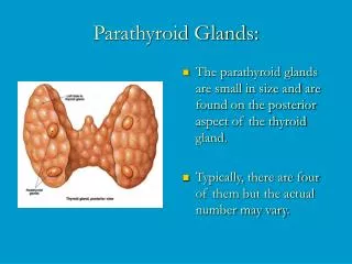
Digestive glands. General outline. small digestive glands. distributed in the wall of digestive tract esophageal glands, gastric glands and intestine glands. large digestive glands. outside the wall of digestive tract salivary glands, liver and pancreas. Large salivary glands.

Digestive glands
E N D
Presentation Transcript
General outline • small digestive glands • distributed in the wall of digestive tract • esophageal glands, gastric glands and • intestine glands • large digestive glands • outside the wall of digestive tract • salivary glands, liver and pancreas
Large salivary glands • Include parotid, submandibular and sublingual glands • are compound tubuloacinar glands • are composed of acini and ducts
Acinus : • acinar epithelium: simple cuboidal or pyramidal cells • myoepithelial cell • basal membrane Structure: Serous acini Mucous acini Mixedacini Be divided into
Serous acini • cytoplasma are deep stained • nucleus are spherical in shape and near the base • secretion is thin, contains salivary amylase and a little mucus
Mucous acini • cytoplasma are light-blue stained • nucleus are flattened ovoid shaped and close to the base • secretion is thick; contains mucoprotein
Mixedacini • consist of above two kinds of cells • demilunes: several serous cells are attached eccentrically to the mucous acini
Ducts: • Intercalated ducts diameter: thinnest wall: simple low cuboidal epithelium
Striated duct (secretory duct) • wall: simple columnar epithelium; the nucleus is near the cell apex; cytoplasm is acidpphilic; has basal striations • EM: the basal striations created by membrane infolding and mitochondia • reabsorbing sodium and excreting potassium; transport water and ions
Interlobular duct Wall: pseudostratified epithelium • Main duct Near its orifice become stratified squamous epithelium
Parotid gland • pure serous gland • longer intercalated duct • secrete 25% of saliva, more salivary amylase, less mucus
Submandibular gland • mixed gland. Serous acini are more than mixed or mucous acini • short intercalated duct, longer striated duct • secrete 70% of saliva, less salivary amylase, more mucus
Sublingual gland • mixed gland, mucous and mixed acini predominant, more demilune • without intercalated duct, obscure striated duct • secrete 5% of saliva, most mucus
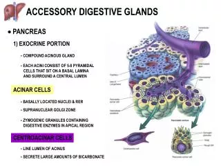
Pancreas Exocrine portion: • the features of the acini • a single layer of pyramidal serous cells surrounded by basal lamina, without myoepithelial cells • centro-acinar cells: the epithelial cells of intercalated duct penetrating into the lumen of the acinus
the feature of ducts • the intercalated duct is long and has branches • no striated duct • main duct: lined by simple columnar epithelium in which a few goblet cells and endocrine cells can be seen
The functions of exocrine portion Secret abundant trypsinogen, chymotrypsinogen, amylase, lipase, sodium bicarbonate and trypsin inhibitor
Endocrine portion (pancreatic islet): • rounded clusters of cells embedded within exocrine pancreatic tissue • are divided into three kinds of cells: A, B, and D cells • fenestrated capillaries are among the cells
Endocrine portion PP cells: pancreatic polypeptide G cells: gastrin Other cells
A cells B cells D cells
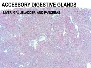
Liver Hepatic plates Sinusoid Central vein Liver lobule Interlobular arteries Interlobular veins Interlobular bile ducts Portal area
Functions of liver: • bile secretion • synthesize: protein, glycogen, cholesterin • detoxification and inactivation • defence • hemopoiesis
Human liver Pork liver
Liver lobule: • hepatic plates: are composed of a single layer of hepatocytes arranged in radial • sinusoid: situated between the hepatic plates, forming a complex network • central vein: occupies the centre of the liver lobule
Hepatocytes • polyhedral in shape, eosinophilic cytoplasm, one or two large rounded nuclei with one or 2 typical nucleoli • EM: Mi, RER, SER, Golgi apparatus, lysosomes, microbodies, inclusions
Mitochondria provide the energy for the hepatocytes • Rough endoplasmic reticulum synthesize some plasma proteins • Golgi apparatus participate in the formation of bile and lipoprotein
Smooth endoplasmic reticulum • synthesize bile, triglyceride and LDL • metabolism of the lipid, hormones and cholerythrin • inactivate steroid hormone • biotransformation of some materials • detoxification of noxious substances
Lysosomes • actively participate the metabolism of hepatocyte and renewal of organelles • play a role in metabolism and transport of cholerythrin • storage of iron
Microbodies detoxification: catalase and peroxidase; reduce the hydrogen peroxide into H2O • Inclusions include glycogen, lipid droplets, pigment etc; These contents vary according to physiologic state of human body
Bile canaliculi • between two adjacent hepatocytes • the membrane of hepatocyte projects to the lumen, forming many microvilli • the cell membranes near the bile canaliculi are firmly bound by junctional complexes
Liver sinusoid • spaces between the hepatic plates • irregular in shape • composed of only one discontinuous layer of fenestrated endothelial cells, no diaphragm, no basement membrane • Kupffer cells are located within the sinusoid cavities
Space of Disse • separates the endothelium from the hepatocytes • contains some reticular fibers and fat-storing cells