Download

1 / 39
400 likes | 745 Views
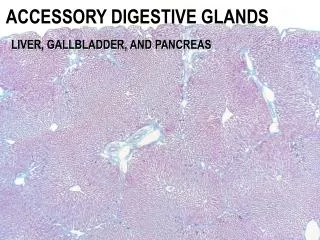
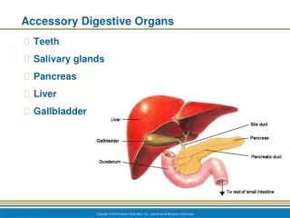
ACCESSORY DIGESTIVE GLANDS. LIVER, GALLBLADDER, AND PANCREAS. ACCESSORY DIGESTIVE GLANDS. TO IDENTIFY THE EXOCRINE AND ENDOCRINE FUNCTIONS OF THE LIVER AND PANCREAS . TO EXAMINE THE HISTOLOGICAL STRUCTURE OF THE LIVER, GALLBLADDER, AND PANCREAS .

E N D
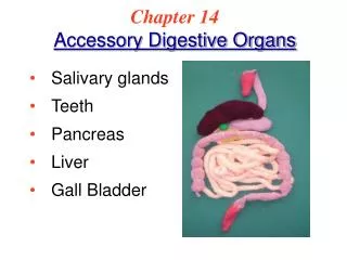
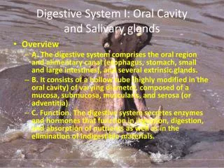
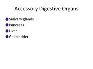
ACCESSORY DIGESTIVE GLANDS LIVER, GALLBLADDER, AND PANCREAS
ACCESSORY DIGESTIVE GLANDS TO IDENTIFY THE EXOCRINE AND ENDOCRINE FUNCTIONS OF THE LIVER AND PANCREAS TO EXAMINE THE HISTOLOGICAL STRUCTURE OF THE LIVER, GALLBLADDER, AND PANCREAS TO CORRELATE STRUCTURE AND FUNCTION OF THESE ACCESSORY DIGESTIVE GLANDS
ACCESSORY DIGESTIVE GLANDS PANCREAS 1) EXOCRINE PORTION 2) ENDOCRINE PORTION
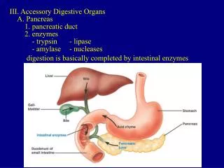
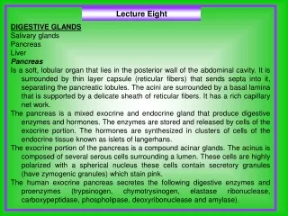
- synthesizes and secretes enzymes via a system of ducts that are essential for digestion in the intestine - synthesizes and secretes hormones into the bloodstream to regulate glucose, lipid, and protein metabolism ACCESSORY DIGESTIVE GLANDS PANCREAS 1) EXOCRINE PORTION 2) ENDOCRINE PORTION
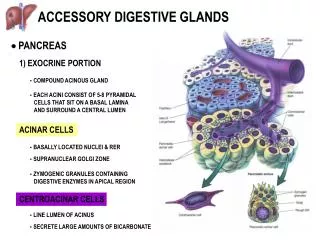
ACINAR CELLS - BASALLY LOCATED NUCLEI & RER - SUPRANUCLEAR GOLGI ZONE - ZYMOGENIC GRANULES CONTAINING DIGESTIVE ENZYMES IN APICAL REGION CENTROACINAR CELLS - LINE LUMEN OF ACINUS - SECRETE LARGE AMOUNTS OF BICARBONATE ACCESSORY DIGESTIVE GLANDS PANCREAS 1) EXOCRINE PORTION - COMPOUND ACINOUS GLAND - EACH ACINI CONSIST OF 5-8 PYRAMIDAL CELLS THAT SIT ON A BASAL LAMINA AND SURROUND A CENTRAL LUMEN
ACCESSORY DIGESTIVE GLANDS PANCREAS 1) EXOCRINE PORTION PANCREAS H&E INTERCALATED DUCTS INTRALOBULAR DUCTS INTERLOBULAR DUCTS MAIN DUCTS
ACCESSORY DIGESTIVE GLANDS PANCREAS 1) EXOCRINE PORTION INTERCALATED DUCTS - lined with simple squamous epithelium INTRALOBULAR DUCTS - lined with simple cuboidal epithelium - size on the order of magnitude of acini INTERLOBULAR DUCTS - lined with simple columnar epithelium - found in fibroconnective tissue septa
ACCESSORY DIGESTIVE GLANDS PANCREAS 1) EXOCRINE PORTION ACINAR CELLS - BASALLY LOCATED NUCLEI & RER - SUPRANUCLEAR GOLGI ZONE - ZYMOGENIC GRANULES CONTAINING DIGESTIVE ENZYMES IN APICAL REGION trypsinogen, amylase, lipase CENTROACINAR CELLS - LINE LUMEN OF ACINUS - SECRETE LARGE AMOUNTS OF BICARBONATE
ACCESSORY DIGESTIVE GLANDS PANCREAS 1) EXOCRINE PORTION INTERCALATED DUCTS PANCREAS H&E
EXO- ENDO- ACCESSORY DIGESTIVE GLANDS PANCREAS 1) EXOCRINE PORTION PANCREAS H&E 2) ENDOCRINE PORTION
ACCESSORY DIGESTIVE GLANDS PANCREAS 2) ENDOCRINE PORTION - PRINCIPLE FUNCTION IS TO SECRETE HORMONES THAT REGULATE BLOOD GLUCOSE LEVELS ISLETS OF LANGERHANS - MASSES OF RICHLY VASCULARIZED ENDOCRINE CELLS SCATTERED THROUGHOUT THE PANCREAS - SEPARATED FROM SURROUNDING ACINAR CELLS BY THIN CAPSULE OF RETICULAR FIBERS - 3 CELL TYPES IN ISLET DISTINGUISHED ONLY VIA SPECIAL STAINS
ACCESSORY DIGESTIVE GLANDS PANCREAS 2) ENDOCRINE PORTION
ACCESSORY DIGESTIVE GLANDS PANCREAS 2) ENDOCRINE PORTION CELL TYPES: 1) ALPHA CELLS (20%) - SECRETE GLUCAGON; RAISES BLOOD GLUCOSE LEVELS 2) BETA CELLS (75%) - SECRETE INSULIN; LOWERS BLOOD GLUCOSE LEVELS 3) DELTA CELLS (5%) - SECRETE SOMATOSTATIN; INHIBITS GLUCAGON AND INSULIN SECRETION
ACCESSORY DIGESTIVE GLANDS LIVER 1) EXOCRINE PORTION - synthesizes and secretes bile via a system of ducts that is essential for digestion in the intestine 2) ENDOCRINE PORTION - synthesizes and secretes numerous plasma proteins into the bloodstream: (albumin, fibrinogen, prothrombin, lipoproteins, etc.)
ACCESSORY DIGESTIVE GLANDS LIVER PORTA HEPATIS PORTAL TRIAD
ACCESSORY DIGESTIVE GLANDS LIVER Classical Liver Lobule - basic functional unit of liver
- central vein at center - hexagonal in shape - short axis: branches of portal triad between 2 classic lobules - portal triad at corners - long axis: between 2 central veins - portal triad at center - triangular in shape - central vein at corners ACCESSORY DIGESTIVE GLANDS LIVER CLASSIC LOBULE PORTAL LOBULE LIVER ACINUS
ACCESSORY DIGESTIVE GLANDS LIVER 1 HEPATOCYTES 2 SINUSOIDS 3 SPACE OF DISSE 4 BILE CANALICULI
ACCESSORY DIGESTIVE GLANDS LIVER 1 HEPATOCYTES 2 SINUSOIDS 3 SPACE OF DISSE - perisinusoidal space between basal surfaces of the endothelial cells and the surfaces of the hepatocytes 4 BILE CANALICULI - small canal beginning the biliary tree formed by apposed grooves in adjacent hepatocytes
ACCESSORY DIGESTIVE GLANDS LIVER
BD HA PV LIVER H&E
ACCESSORY DIGESTIVE GLANDS LIVER BILE DUCTS - lined with simple cuboidal epithelium
ACCESSORY DIGESTIVE GLANDS LIVER LIVER H&E 1 HEPATOCYTES 2 SINUSOIDS
ACCESSORY DIGESTIVE GLANDS LIVER LIVER RETICULAR FIBER MESHWORK
ACCESSORY DIGESTIVE GLANDS LIVER RETICULAR FIBER MESHWORK KUPFFER CELLS - phagocytic cell, liver macrophage
ACCESSORY DIGESTIVE GLANDS LIVER LIVER H&E KUPFFER CELLS CV
ACCESSORY DIGESTIVE GLANDS LIVER LIVER H&E KUPFFER CELLS
ACCESSORY DIGESTIVE GLANDS LIVER 3-D APPRECIATION FOR HISTOLOGICAL STRUCTURE OBSERVED IN 2-D
ACCESSORY DIGESTIVE GLANDS LIVER 3-D LOOK AT THE BILIARY SYSTEM
ACCESSORY DIGESTIVE GLANDS LIVER H&E LIVER SITE OF HEMOPOEISIS DURING EARLY GESTATION
ACCESSORY DIGESTIVE GLANDS LIVER H&E LIVER SITE OF HEMOPOEISIS DURING EARLY GESTATION
HEPATOCYTE BILE CANALICULI RT & LT HEPATIC DUCTS COMMON BILE DUCT ACCESSORY DIGESTIVE GLANDS GALLBLADDER - LOCATION FOR CONCENTRATION AND STORAGE OF BILE ROUTE OF BILE FILLING OF GALLBLADDER
ACCESSORY DIGESTIVE GLANDS GALLBLADDER THREE LAYERS: 1) MUCOSA - lined with tall columnar epithelium and underlying basal lamina & lamina propria - mucosa highly folded and irregular - NO MUSCULARIS MUCOSA OR SUBMUCOSA - 2) MUSCULARIS EXTERNA - layers of smooth muscle with irregular orientation 3) ADVENTITIA or SEROSA
ACCESSORY DIGESTIVE GLANDS GALLBLADDER EPITHELIUM: - tall columnar epithelium and underlying basal lamina & lamina propria - basally located nuclei - fine microvilli border