Download
1 / 25
260 likes | 298 Views
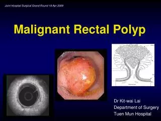
Malignant colonic polyp: endoscopic treatment updates. CHAN Ka-man, Fiona Kwong Wah Hospital Joint Hospital Surgical Grand Round 18 th April, 2015. Prevalence. Markowitz AJ. CA Cancer J Clin 1997;47:93-112 Nusko G. Endoscopy 1997;29: 626-631 Williams AR. Gut 1982;23:835-42.

E N D
Malignant colonic polyp: endoscopic treatment updates CHAN Ka-man, Fiona Kwong Wah Hospital Joint Hospital Surgical Grand Round 18th April, 2015
Prevalence Markowitz AJ. CA Cancer J Clin 1997;47:93-112 Nusko G. Endoscopy 1997;29: 626-631 Williams AR. Gut 1982;23:835-42 • Screen detected adenoma • 21-58% from 50-70 years • Malignant polyps in endoscopically removed polyps • 0.2-11%
Superficial neoplastic lesion Wolff WI. Annals of Surgery 1975;182:516-525 Japanese Society for Cancer of the Colon and Rectum. 2009 World Health Organization classification of tumors. 2010. pp. 104–109 • Malignant colonic polyp • Neoplasm that penetrates the muscularis mucosae into submucosa • Carcinoma in-situ/High-grade intraepithelial neoplasia • Neoplasm that are confined to the epithelium or invade the lamina propria alone and lack invasion through the muscularis mucosae
Why is endoscopic treatment feasible? • Risk of lymph node metastasis in Tis is negligible • Risk of lymph node metastasis in submucosal lesion • Risk 6-12% in general • Pedunculated lesions • Rate of lymph node metastasis was 0% in head invasion cases and stalk invasion cases with SM depth <3000 µm if lymphatic invasion was negative. • Non-pedunculated lesions • Rate of lymph node metastasis was also 0% if SM depth was <1000 µm.
Classification Paris Classification Japan Classification Gastrointest Endosc 2003; 58(Suppl. 6): S3–43 Japanese Classification of Colorectal Carcinoma. 1997
Lateral spreading tumour (LST) • Neoplasm with horizontal extending growth pattern • >10mm • Granular type (LST-G) • Non-granular type (LST-NG) • High possibility of deep submucosal invasion 14% versus 7% in glandular type (p<0.01) • 30-56% have multifocal invasion Japanese Classification of Colorectal Carcinoma. 1997
Endoscopic treatment options Williams. Colorectal Disease 2013;15:1–38
Chromoendoscopy Narrow band imaging Exclusion of lesion for endoscopic treatment Kudo. Gastrointest Endosc 1996;44:8-14 Sano. Digest Endosc, Vol. 18.S44–51
Endoscopic treatment • Snare polypectomy • Endoscopic mucosal resection (EMR) • Endoscopic submucosal dissection(ESD)
Piecemeal EMR John Hopkins colon cancer center
Efficacy of EMR Wada. Stomach Intestine 2013;48:134–44 Walsh. Gastrointest Endosc 1992;38:303–9 Saito. Gastrointest Endosc Clin N Am 2010;20:515–24 Jin. Cancer Therapy. Vol. 7. pp. 27-30 • En bloc resection: 66.5–80% when the tumor sizes were <20 mm • When the tumor sizes were ≥20 mm, the en bloc resection rate significantly decrease to 20-48% • Local recurrence • 3% en bloc resection • 20% piecemeal resection
Endoscopic submucosal dissection (ESD) Kōdansha. Understanding ESDs: A Procedure for Treating Cancer Without Major Surgery. 2011
Efficacy of ESD Tanaka S. Dig Endosc 2012; 24(Suppl 1):73–79 Saito. Gastrointest Endosc 2010;72:1217–1225 Puli SR. Ann Surg Oncol 2009;16:2147-2151 • Meta-analysis of ESD of 1314 large flat polyps • En-bloc resection rates 88%-90.5% • Histological R0 resection rate 76.9%-89% • Local tumor recurrence 1.9%
ESD vs. EMR Nakajima. Surg Endosc 2013 Saito. Surg Endosc 2010;24:343–352 Larger resected specimens (37 mm vs. 28mm; p=0.0006) Higher en-bloc resection rate(94.5% vs. 56.9%; p<0.01) Less recurrences (2% vs. 14%; p<0.0001) Longer procedure time (108-129 min vs. 18-29 min; p<0.0001) Higher perforation rate (6.2% vs. 1.3%)
ESD versus laparoscopic colectomy Kiriyama S. Endoscopy 2012; 44:1024–1030 Nakamura. SurgEndosc 2015;29:596-606 Limited comparative data Shorter procedure time (95 vs. 185 mins; p<0.001) Shorter hospital stay (5 vs. 10days; p<0.001) Less analgesic requirement Early resumption of diet and mobility The 3-year overall survival rate exceeded 99% in both the ESD and LAC groups
Difficulty in ESD Uroka. Journal of Gastroenterology and Hepatology (2013) 406–414 • Anatomical difficulties • Longer length, narrower lumen, extensive flexion and thinner walls • Steep learning curve • Animal models • 20 gastric ESD → rectal ESD → colon ESD • Complication • Perforation rate 4-10% • Bleeding rate 0.7-2.4%
Curative endoscopic resection • Lateral and vertical margins of the specimen were free • Submucosal invasion less than 1,000 μm • No lymphovascular involvement • No poorly differentiated component • Tumor budding grade 1 (low grade) Kitajima. J Gastroenterol 2004; 39:534–543
Endoscopic surveillance Repici .Dis Colon Rectum 2009; 52: 1502–15 Detection of recurrence Metachronous adenoma and early carcinoma were detected in 54.8% and in 11.9% of surveillance endoscopy No evidence-based consensus First surveillance at 3-6 months, then regular surveillance in 3-5 years
Conclusion Malignant colonic polyps can be managed by endoscopic resection ESD enables en-bloc resection of large superficial tumours Regular surveillance aids detection of recurrence which can be managed endoscopically
Reference Wolff WI, Shinya H. Definitive treatment of “malignant” polyps of the colon. Annals of Surgery. 1975;182(4):516-525. Japanese Society for Cancer of the Colon and Rectum, editor. Japanese Classification of Colorectal Carcinoma. 2nd ed. Tokyo: Kanehara & Co., Ltd; 2009 Kitajima K, Fujimori T, Fujii S et al. Correlations between lymph node metastasis and depth of submucosal invasion in submucosal invasive colorectal carcinoma: a Japanese collaborative study. J. Gastroenterol. 2004; 39: 534–43. Participants in the Paris Workshop. The Paris endoscopic classification of superficial neoplastic lesions: esophagus, stomach, and colon: November 30 to December 1, 2002. Gastrointest Endosc 2003; 58(Suppl. 6): S3–43. Oka S, Tanaka S, Kaneko I et al. Conditions of curability after endoscopic treatment for colorectal carcinoma with submucosal invasion: Assessments of prognosis in cases with submucosal invasive carcinoma resected endoscopically. Stomach Intestine 2004; 39: 1731–43.
Polyp morphology relation to size and risk of submucosal invasion The Paris endoscopic classification of superficial neoplastic lesions. Gastrointest Endosc 2003; 58(Suppl. 6): S3–43