Download

1 / 39
440 likes | 1.88k Views
Treatment of Early Malignant Rectal Polyp . Dr KP Tsui Department of Surgery Tseung Kwan O Hospital. Malignant Rectal Polyp. Polyps with cancer cells invading the muscularis mucosa Invasion limited to submucosa T1 lesion .

E N D
Treatment of Early Malignant Rectal Polyp Dr KP Tsui Department of Surgery Tseung Kwan O Hospital
Malignant Rectal Polyp • Polyps with cancer cells invading the muscularis mucosa • Invasion limited to submucosa • T1 lesion
Incidence of malignant colorectal polyps as a proportion of all adenomas removed varies between 2.6 and 9.7%. • Average 4.7% Sobin L, Wittekind C (eds). TNM classification of Malignant Tumours (6th Edition). Wiler-Liss: New York, 2002.
Size most important determinant factor determining risk of malignant transformation within a polyp • > 1 cm: 38.5% • > 42 mm: 78.9% Tytherleigh et al. BJS 2008;95:409-423
Villous adenomas have highest risk of malignancy at 29.8% • Tubular adenomas have lowest at 3.9% Tytherleigh et al. BJS 2008;95:409-423
Treatment • Staging • Histological Assessment
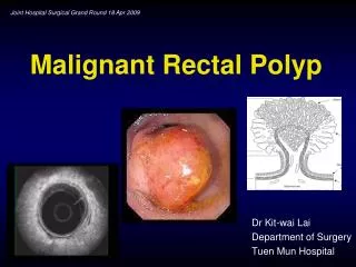
Colonoscopy: 2 cm rectal polyp (5 cm from anal verge) Biopsy: adenocarcinoma Clinical Scenario 1
Endorectal ultrasound • Best method to differentiate between T1 and T2 lesion • T stage N stage Accuracy: 90 % Accuracy: 80% Sensitivity : 85% Sensitivity: 70% Specificity: 95% Specificity: 80% Bretagnol et al. Dis Colon Rectum 2007;50:523-533
Can assess residual tumor after polypectomy • Follow up after local excision Hernandez De Andaetal. Dis Colon Rectum 2004; 47: 818–824
Limitations • Operator dependent • Upper rectal lesions • Tumor stenosis • Peritumoral fibrosis and inflammatory tissue • Effect of radiotherapy or hemorrhage after biopsy
Pelvic MRI • Overall T stage accuracy 59-95% • T1,2 lesion (vs ERUS) - Similar sensitivities - Lower specificity (69%) • N stage - Comparable to EUS • Can evaluate entire pelvis Bretagnol et al. Dis Colon Rectum 2007;50:523-533 Tytherleigh et al. BJS 2008;95:409-423
CT abdomen + pelvis • Distant metastases • Low accuracy for T staging, 52 – 94% and N stage, 54-70% Alexandre Jin Bok Audi Chang et al. Journal of Surgical Education; Vol 65: Number 1 Bretagnol et al. Dis Colon Rectum 2007;50:523-533
PET • Limited role for local and regional staging • Sensitivities for lymph node metastases 22-29% Abdel-Nabi H, Doerr RJ, Lamonica DM, et al. Radiology. 1998;206:755-760
Surgical Options Local excision vs Radical Surgery Park’s per anal excision Abominoperineal resection TEM Total Mesorectal Excision Anterior resection
Local Excision • Opportunity of cure with less detriment • Sphincter preservation • Less morbidity and mortality • Less sexual or urinary dysfunction
Park’s per anal excision • Aid of anal retractors • 6-10 cm of anal margin • Full thickness excision • At least 1 cm margin • Defect usually closed with absorbable sutures
Transanal endoscopic microsurgery • Rectoscope • Usually below peritoneal reflection • Full thickness excision • Excision margin of 1 cm • Difficult for lesions within 6 cm
Complications • Overall rate 6-31% • Postoperative hemorrhage 1-13% • Perforation 0-9% • Suture line dehiscence • Perirectal abscess • Rectal stenoses Hiroko Kunitake, et al. Perm J 2012 Spring;16(2):45-50
Local Excision Vs Radical Surgery
Generally accepted that local excision, by either endoscopic polypectomy or transanal surgery is adequate treatment for low risk ERC Tytherleigh et al. BJS 2008;95:409-423
Poorly differentiated carcinoma: 50% risk of lymph node metastasis Coverlizza S, Risio M, Ferrari A, Fenoglio-Preiser CM, Rossini FP. Cancer 1989;64:1937-47 • Lymphovascular invasion, sm3 invasion, undifferentiated carcinomas have significant risks of LN metastases. Nascimbeni et al. Dis Colon Rectum 2002;45:200-206
Des. • Depth of invasion was found to be best estimate of the probability of regional LN metastasis Bretagnol et al. Dis Colon Rectum 2007;50:523-533 • Rate of lymph node metastasis Sm1 1-3% Sm2 8% Sm3 23% Nascimbeni et al. Dis Colon Rectum 2002;45:200-206
Optimal choice of surgery • The role of local excision as a curative procedure has been questioned due to inferior outcome in some long term follow up series. Alexandre Jin Bok Audi, MD, et al. Journal of Surgical Education; Vol 65: Number 1 (2008)
Alexandre Jin Bok Audi, MD, et al. Journal of Surgical Education; Vol 65: Number 1 (2008)
Most literature data are based on case reports or small series with no standard criteria for patient selection
Adjuvant chemoradiotherapy • May be beneficial • Recommended for high risk T1 lesions, assuming further surgery is not an option Tytherleigh et al. BJS 2008;95:409-423
Limitations • Most retrospective studies • Lack of controlled data • No defined protocol for chemotherapy
Salvage surgery • Between 56 and 100% of recurrence suitable for salvage surgery • May not offer same outcomes as initial treatment • Should not be delayed in case of recurrence Tytherleigh et al. BJS 2008;95:409-423
Clinical Scenario 2 • Colonoscopic polypectomy of rectal polyp • Pathology: adenocarcinoma
Pathology No High Risks Features Haggitt level 1,2,3 Kikuchi Sm1 High Risks Features Sm3 (Sm2) Grade lymphovascular ERUS MRI CT LN- LN+ Margin involvement Yes Histological assessment not adequate No Local Excision No Yes Follow up Radical Surgery High Risks Features
Follow up • Digital rectal exam + Endoscopy + CEA First 3 years: every 3 months Next 2 years: every 6 months Then annually • Endorectal ultrasound should be performed at every outpatient session Mellgren et al. Dis Colon Rectum 2000; 43: 1064–1071 NCCN guideline
Summary • Local excision Recommended for low risk T1 Sm1 lesion • Radical surgery For high risk T1 lesion Adjuvant therapy if further surgery is not an option
Recurrence Diagnose early for salvage surgery • Follow up Endoscopic surveillance of rectum and scar