Download
1 / 34
400 likes | 1.08k Views
COLONIC DIVERTICULAR DISEASE. INCIDENCE & EPIDEMIOLOGY. * US Census Bureau, International Data Base, 2004 ( the extrapolations for Diverticular Disease are only estimates and may have limited relevance to the actual incidence of Diverticular Disease in any region).

E N D
* US Census Bureau, International Data Base, 2004 ( the extrapolations for Diverticular Disease are only estimates and may have limited relevance to the actual incidence of Diverticular Disease in any region)
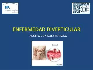
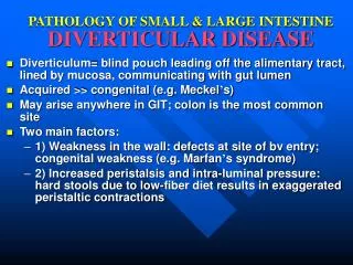
TYPES • FALSE DIVERTICULA • involves only protrusion of the mucosa through the muscularispropria of the colon • most common • TRUE DIVERTICULA • a saclike herniation of the entire bowel wall
PATHOPHYSIOLOGY Protrusion occurs at the point where the NUTRIENT ARTERY or VASA RECTI penetrates through the muscularispropria Break in the integrity of the colonic wall Compression or erosion PERFORATION BLEEDING
PATHOPHYSIOLOGY • commonly affect the SIGMOID COLON due to: • Relative high pressure zone within the muscular sigmoid colin. • Higher amplitude contractions combined with constipated, high fat content stool within the sigmoid lumen results in the creation of these diverticula • Related to retention of particulate material within the diverticular sac and formation of fecalith
Diverticular bleeding Presentation, Evaluation, and Management
Diverticular Bleeding • Hemorrhage from a colonic diverticulum is the most common cause of hematochezia in patients >60 years. • Only 20% of patients with diverticulosis will have GI bleeding. • Most bleeds are self-limited and stop spontaneously with bowel rest. • Lifetime risk of rebleeding: 25%
Diverticular Bleeding • Colonoscopy • To localize the bleeding • May be both diagnostic and therapeutic in the management of mild to moderate diverticular bleeding • Angiography • Management of massive bleeding in a stable patient • Mesenteric angiography can localize the bleeding site and occlude the bleeding vessel successfully with a coil in 80% of the cases • Follow up: Repetitive colonoscopy to look for evidence of colonic ischemia • Segmental resection of the colon • To eliminate risk of further bleeding • In patients on chronic blood thinners
Diverticular Bleeding • Highly selective coil embolization • Rate of colonic ischemia: <10% • Risk of acute rebleeding: <25% • Selective infusion of vasopressin • To stop hemorrhage • Complications: MI, intestinal ischemia • Recurrence of bleeding in 50% of patients once infusion is stopped
Diverticular Bleeding • Surgery • Indications: if patient is unstable or has had a 6-unit bleed within 24 h • Total abdominal colectomy • Patients with presumed bleeding from diverticular disease requiring emergent surgery without localization • Rationale: Colonic diverticulosis is more often seen from the R colon • Surgical resection with primary anastomosis • In patients without severe comorbidities
Diverticulitis Presentation, Evaluation, and Management
Diverticulitis • Diverticular perforation • Generalized peritonitis in <25% of cases • (+) Abdominal distention • Giant diverticulum of the sigmoid • (+) Air fluid level in the LLQ on plain abdominal film • Mx: resection to avoid impending perforation
Diverticulitis • Diagnosis is best made on CT.
Diverticulitis • Suspected diverticulitis that does not meet CT criteria or is not associated with a leukocytosis or fever is not diverticular disease • Conditions that mimic diverticular disease: • IBS • Ovarian cyst • Endometriosis • Acute appendicitis • PID
Diverticulitis • Barium enema or colonoscopy • Should be performed ~6 weeks after an attack of diverticular disease • A sigmoid malignancy can masquerade as diverticular disease. • Should not be performed in the acute setting • Higher risk of colonic perforation associated with insufflation or insertion of barium-based contrast material under pressure.
Diverticulitis • Complicated diverticular disease • Diverticular disease associated with an abscess or perforation, and less commonly with a fistula. • With fistula formation • Common locations include cutaneous, vaginal or vesicle fistulae • Present with either passage of stool through skin or vagina, or pneumaturia • Colovaginal fistulae: more common in women who have undergone hysterectomy
Medical Management of Diverticular Disease • Asymptomatic • Diet alterations – fiber-enriched diet, including 30g of fiber/day • Supplementary fiber products: Metamucil, Fibercon, Citrucel • Avoid nuts and popcorn – may obstruct the lumen of the diverticulum
Medical Management of Diverticular Disease • Symptomatic • Radiographic and hematologic confirmation of inflammation and infection within the colon • Treated initially with antibiotics and bowel rest • TMP-SMX or ciprofloxacin and metronidazole • (+) Ampicillin – for nonresponders • Alternative: IV piperacillin or oral penicillin/clavulinic acid • Usual course: 7-10 days • Rifixamin + fiber – less frequent recurrent symptoms from uncomplicated diverticular disease • Limited diet until pain resolves • Medical therapy can be continued beyond 2 attacks without an increased risk of perforation requiring a colostomy, especially in those >50 years.
Surgical Management ofDiverticular Disease • In patients who are low risk (ASA I and II) who have had at least 2 documented attacks requiring hospitalization or those who do not rapidly improve on medical therapy • Younger patients – more aggressive form of disease • Waiting beyond two attacks is not recommended. • In all low surgical risk patients with complicated diverticular disease
Surgical Management ofDiverticular Disease • Goals of Surgical Management • Control sepsis • Eliminate complications such as fistula or obstruction • Remove diseased colonic segment • Restore intestinal continuity
Surgical Management ofDiverticular Disease • Open or laparoscopic sigmoid resection – current option of uncomplicated diverticular disease • Benefits of laparoscopic over open resection: • Early discharge (by at least 1 day) • Less narcotic use • Earlier return to work • Benefits of open over laparoscopic resection: • Shorter operative procedure • Less costly • Complication rates are similar.
Surgical Management ofComplicated Diverticular Disease • Proximal diversion of the fecal stream with an ileostomy or colostomy and sutured omental patch with drainage • Resection with colostomy and mucus fistula or closure of distal bowel with formation of a Hartmann’s pouch • Resection with anastomosis (coloproctostomy) • Resection with anastomosis and diversion (coloproctostomy with loop ileostomy or colostomy)
Surgical Management ofDiverticular Diseases • Hinchey Stages I and II • Percutaneous drainage followed by resection with anastomosis about 6 weeks later • Percutaneous drainage • For abscesses ≥ 5 cm with a well-defined wall that is accessible • If <5cm, may resolve with antibiotics alone • Contraindications to percutaneous drainage: • No percutaneous access route • Pneumoperitoneum • Fecal peritonitis
Surgical Management ofDiverticular Diseases • Hinchey Stages I and II • If patients develop generalized peritonitis Hartmann’s procedure • Nonoperative therapy – 20% recurrence rate at 2 years in patients with Hinchey Stage I disease • 80% of patients with Hinchey Stage II required surgical resection for recurrent symptoms.
Surgical Management ofDiverticular Diseases • Hinchey Stage III • Hartman’s procedure or with primary anastomosis and proximal diversion • If patient has significant comorbidities: intraoperative peritoneal lavage (irrigation), omental patch to the oversewn perforation, and proximal diversion of the fecal stream with either an ileostomy or transverse colostomy can be performed • Hinchey Stage IV • No anastomosis of any type should be attempted.
Recurrent Symptoms inDiverticular Disease • Occurs in 10% of patients. • Recurrence develops in patients following inadequate surgical resection. • A retained segment of diseased rectosigmoid colon is associated with twice the incidence of recurrence. • IBS – may also cause recurrence of initial symptoms • Patients undergoing surgical resection for presumed diverticulitis and symptoms consistent with IBS have functionally poorer outcomes.