Download
1 / 57
570 likes | 738 Views
Learn about the functions, structure, and organs of the digestive system. Explore the processes involved in digestion and absorption of nutrients, as well as the role of accessory organs. Discover the layers of the digestive tract and the features of the peritoneum. Gain insights into the mouth anatomy, teeth structure, and salivary glands' functions. Understand the functions of the esophagus and stomach in mechanical and chemical digestion.

E N D
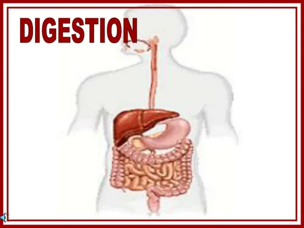
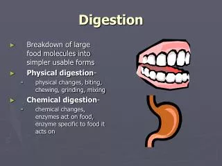
Digestion I. Functions of the digestive system 1. Take in food. 2. Break down food. 3. Absorb digested molecules. 4. Provide nutrients. 5. Eliminate wastes.
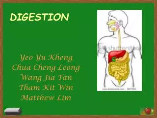
II. Digestive Structure & Function A. Organization 1. Alimentary Canal (GI tract). a. 30 ft b. Digest food c. Absorb nutrients d. Eliminate wastes 2. Accessory Organs a. Located within or outside the GI tract.
B. Digestive Processes 1. Ingestion 2. Mechanical Digestion 3. Propulsion 4. Chemical Digestion 5. Absorption 6. Defecation
Review • State the functions of the digestive system. • Name & describe the 6 processes of the digestive system.
III. Features of the Digestive System A. Peritoneum 1. Parietal –serous membrane that lines the abdominal pelvic walls. 2. Visceral (serosa) – lines the organs. 3. Peritoneal cavity – Space within the abdominal pelvic cavity. 4. Falciform Ligament supportive ligament for liver. 5. Mesentery- connective tissue that holds many abdominal organs in place. 6. Lesser Omentum- mesentery that connects the lesser curvature of the stomach to the liver and diaphragm. 7. Greater Omentum (mesentery proper) – mesentery connecting the greater curvature of the stomach to the transverse colon.
8. The greater omentum is usually long, double fold mesentery that extends inferiorly and loops back to the colon creating a cavity known as the Omental bursa. 9. Greater omentum usually fills with fat depoists. 10. Retroperitoneal- abdominal organs that lie behind the peritoneum.
B. Wall Structure of the Alimentary Canal 1. Mucosa – Mucus membrane that lines the lumen. 2. Submucosa – Loose connective tissue (blood & lymph tissues). 3. Muscularis – muscle (circular & longitudinal fiber). 4. Serosa – Visceral peritoneum, outer covering.
Review • What is the functions of the mesenteries, greater & lesser omentum? • What are the layers of the digestive tract?
IV. Digestive Organs of the Alimentary Canal. A. Mouth & Accessory Organs 1. Lips & Checks 2. Palate 3. Tongue – large muscular organ. frenulum-attachs tongue to floor of mouth. 4. Teeth
a. Deciduous 20 by 2 yrs. b. Permanent 32 between ,17-25 yrs. 1. Incisors – chisel – cutting 2. Canines – cone shape – tearing 3. Premolars 4. Molars – broad & flat - grinding
c. Structure 1. Crown – visible part of the tooth. * Enamel – protection, hardest substance of the body. *Cells die shortly after tooth break through, incapable of repair. 2. Neck – between two regions. 3. Root – below gums. 4. Dentin – slightly harder than bone, surrounds pulp cavity. 5. Pulp-Soft connective tissue (blood & nerve).
Review • List the functions of the lips, cheeks, & tongue. • What are deciduous and permanent teeth? Name the different kinds of teeth? • Describe the parts of a tooth. What are dentin, enamel, & palate?
6. Salivary Glands a. Saliva – 99.5% water, .5% solutes 1. Liquid medium for dissolving food particles. 2. Lubrication for swallowing. 3. Enzymes – Lysozyme – destroys bacteria. Salivary Amalyase – begins chemical digestion of carbohydrates. b. Glands 1. Parotid – Largest in front of ears. 2. Submandibular – floor of mouth for mucus. 3. Sublingual – under tongue, for mucus.
c. Digestion in Mouth 1. BEGINS IN THE MOUTH. 2. Mastication – Chewing. 3. Bolus – compact mass of food & salvia. B. Pharynx & Esophagus 1. Transports food from the mouth to the stomach WITHOUT further processing a. Nasopharynx b. Oropharynx c. Laryngopharynx 2. Deglutition a. Voluntary – bouls moves to the back of the mouth b. Pharyngeal- reflex begins with the evevation of the soft palate, then pharyngeal muscles contract to force food through the pharynx. Epiglottis is tipped posteriorly. c. Esophageal- swallowing by peristaltic waves
Review • What is the function of saliva? • Name the salivary glands. • What is the job of the esophagus?
C. Esophagus 1. Muscular tube 2. 10 inches long 3. Collapsed when not propelling food 4. Behind trachea 5. Esophageal hiatus – opening of esophagus 6. Esophageal sphincters a upper b. Lower esophageal sphincter – valve that prevents upward movement. heartburn – weak & permits leakage of stomach juices which irritation to esophagus. D. Stomach – Mechanical & Chemical Digestion 1. Stomach a. Pouch – 10 inches long b. Rugae – deep folds in inner lining c. Greater Curvature – Convex d. Lesser Curvature – Concave
e. Four Regions 1. Cardia – Opening that receives food. 2. Fundus – Expanded regions above cardia, temporary holding regions. 3. Body – Main part. 4. Pylorus – Narrow inferior region f. Sphincter – Keep content within stomach 1. Cardiac – Esophagus & Stomach 2. Plyorus – Stomach & small intestines
2. Stomach Wall a. Four basic layers 1. Mucosa -gastric pits -gastric glands Parietal cell – HCL, Intrinsic factor Chief Cell – Pepsinogen -> pepsin Mucus Cells – Mucus -Gastric Juices 2-3 L/day 2. Sub-mucosa 3. Muscularis Additional layers that mix & churn 4. Serosa
3. Function of Stomach a. Mechanical Digestion – mixing & churning b. Chemical Digestion – Gastric juices 1. Chief Cell – Pepsinogen (inactive to prevent it from digestion of own cells) 2. Parietal Cell – HCL which activates pepsinogen to pepsin. 3. Pepsin breaks down nearly all protein. 4. Mucus – Coats to prevent digestion of stomach. -gastric ulcers
c. Absorption 1. Salts 4. Asprin 2. Glucose 5. Lipid Soluble drugs 3. Alcohol 6. Water d. Propulsion – Peristalsis 1. Chyme- small food particles & gastric juices. 2. Mixing waves- weak contraction which mixes food with stomach secretions. 3. Peristalic waves- stronger contractions that force chyme toward and through the pyloric sphincter. e. Intrinsic Factor Parietal & Chief Cells – aids in Vitamin B12 absorption by small intestines, which is important in RBC production.
Review • Describe the parts of the stomach. • How are the stomach muscles different from those in the esophagus?
f. Regulation of Stomach Function 1. Beginning of Digestion -Primary by involuntary control center of the brain & hormones – Positive Feedback Brain (hypothalmus) Medulla Cephalic Phase - Smell, think, see, hear Gastric phase -Gastric Glands Gastirc Juices Continued Reflex (Stretching of stomach by food) 2. Ending of Digestion Intestinal phase -Presence of acid in upper small intestine triggers nerve reflex to stimulate contraction of the pyloric valve, which prevents more acid from entering .-Secretin & Cholecystokinin (CCK) intestinal hormones. Inhibits stomach peristalsis & stimulate pancreatic enzyme & bile into small intestines.
3. Regulation a. Controlled by the involuntary brain & hormones 1. Secretnin -Released into the bloodstream because of the HCL in the small intestines. -inhibits the gastric secretion. -Causes pancreas to release bicabonate ions to neutralize the pH. 2. CCK -Release in response to fats & proteins into the small intestines. -Cause the pancreas to release pancreatic juices. -Digestion to release pancreatic juices. -Inhibits gastric juices & peristalsis.
4. Small Intestines A. Structure 1. Longest segment of the GI tract (20 ft). 2. 1 inch in diameter. 3. Three segments -Duodenum- receives chyme from the stomach,10 in long. -Jejunum – 8ft -Ileum- 12 ft – unites the small & large intestines - Ileocecal Valve
B. Wall of intestines – Same four layers with modification - increase Sa by 600 X’s circular folds mucosa & submucosa villi lacteal – lymphatic vessel microvilli
C Layers 1. Mucosa a. cells -absorptive – have microvilli, produce digestive enzymes, absorb digested food. - goblet – produce mucus. - endocrine – produce regulatory hormones. - granular (Paneth’s cell)- help protect from bacterial infection. b. glands Intestinal glands – secretes water & mucus. Duodenal glands – produce mucus. 2. Submucosa Bruner’s glands – only in walls of duodenum for neutralizing. (Payer’s patches) – fights off infection. 3. Muscularis 4. Serosa
C. Function 1. Completes chemical digestion a. Pancreatic enzymes – pancreatic duct b. Bile – common bile duct - peptidases- proteins - disaccharides- disaccharides into monosaccharides. (maltose & isomaltose) 2. Absorption – main site of nutrient absorption (duodenum & jejunum). 3. Propulsion – mix & move contents toward the large intestines. Peristaltic contraction- through the intestine. Segmental contraction – mixing in short segments a. Diarrhea – too fast b. Constipation – too slow
Review • 1. What secretions complete the chemical digestion of food particles? • 2. What nutrients can be absorbed by the intestinal walls? • 3. How are the layers of the small intestine modified for their function? • 4. What are circular folds, villi, & microvilli in the small intestine? What are their functions?
5. Large Intestines - Absorption of water - Process of defecation A. Structure 1. 5 ft, 3in in diameter 2. 4 segments. a. Cecum- Receives material from the ileum. Short pouch like segment below ileoceal valve. Vermoform appendix – appendicitis. b. Colon- Main absorption site Ascending Transverse Descending Sigmoid
B. Rectum – Storage of fecal material C. Anal canal – Opens to the exterior. Internal sphincter – involuntary- smooth. External- voluntary - skeletal
D. Wall of intestine 1. Same four layers with modification. -Mucosa – Lack villi, has deep folds, large number of mucus secreting cells to bind contents together & for easier defecation. -Submucosa- deep folds -Muscularis – three bands taenai coli form pouches called – haistra. -Anal Canal – Parallel ridges to reduce friction during defecation.
2. Function a. Mucosa absorbs much of the remaining water & electrolytes in chyme. b. Chyme harden – feces 75% water & 25% indigestible wastes. c. Color is a result of bile altered by bacteria. 1. Most there than anywhere else in the body. 2. Produces vitamin K 3. metabolism produces gases, CO2, Methane, hydrogen sulfide. 4. Most of gas is swallowed air. 3. Defecation or Elimination a. Propulsion (peristalsis of feces through the large intestines (18-24 hrs) for colon. mass movements- 8-12 hrs , moves contents 20 cm towards anus. b. Stores until the rectum is full c. Once full, nerve receptors in rectal wall stimulate defecation reflex. parasympathetic reflex
Review • 1. What are the divisions of the large intestines? • 2. What is the function of the large intestines? • 3. How is chyme converted to feces? • 4. Describe the defecation reflex.
V. Accessory Organs A. Pancreas – Wide range of enzymes 1. Structure a. Head Body Tail b. Acini – exocrine secretory cells. Pancreatic juices into the pancreatic ducts & fuses with the common bile duct. c. Islets of Langerhans – Produces insulin & glucagon 2. Function a. 1500ml/day b. Pancreatic Amylase – Carbohydrates c. Tyspin – Protein d. Chyotrypsin – Protein e. Pancreatic Lipase – Lipids f. Nucleases – breaks down DNA & RNA
B. Liver – Largest internal organ of the body at 1.36 Kg. 1. Structure a. Lobes or sections 1. Right – large 2. Left – small b. Caudate and quadrate– smaller sub divisions of the lobes. c. Porta – gateway to the liver d. Hepatic artery- oxygen rich blood to the liver e. Hepatic veins – oxygen poor blood from the liver to the vena cava. f. Hepatic portal vein – oxygen poor blood but high in nutrients to the liver. g. Lobules-smaller divisions . h. Portal triads three structures 1. hepatic artery 2. hepatic portal vein 3. hepatic duct i. Hepatic cords j. Hepatic sinusoids k. Hepatocytes
k. (cont.)Hepatocytes – Liver cells arranged in columns around a central vein. l. Kupffer cells – Phagocytic cells that remove bacteria from the blood that came from the digestive tract. m. Bile Canaliculi- Secrete bile. n. right hepatic duct o. cystic duct p. Common bile duct q. Duodenal papilla opening in small intestines from the common bile duct and the pancreatic duct.
2. Function a. Secretion of Bile, 700 ml/day -Emulsification of fats. -Absorption of vitamin A,D,E, & K. -bilirubin-bile pigments b. Metabolism of lipids -Lipoproteins – package of fat for transport & storage. -HDL’s – transports cholesterol to liver – beneficial in repair, growth, but tends to accumulate in the body spaces when not used. -LDL’s – Transport cholesterol to cells-production of hormones, harmful-atheroscerosis. -VLDL’s c. Stores glycogen, vitamins A, D, B12 d. Detoxifies harmful substances in the blood stream – alcohol.
3. Control of Bile secretion & release a. Secretin – liver b. Cholecystokinin – gall bladder c. 90 % of bile is reabsorbed in the ileum
V. Digestion, Absorption and Transport A. Carbohydrates 1. cotransport and active transport
B. Lipids 1. simple diffusion 2. Chyle – lymph that contains high amount of absorbed lipids.