Download

1 / 63
650 likes | 686 Views
Explore the structural differences between prokaryotic and eukaryotic cells, including their shapes, arrangements, cell walls, flagella, and motility mechanisms. Understand the significance of Gram staining, cell wall damage, plasma membrane structure, and membrane transport processes.

E N D
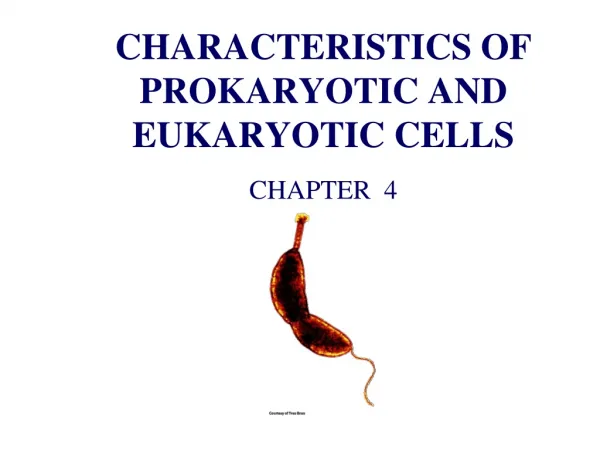
4 Functional Anatomy of Prokaryotic and Eukaryotic Cells
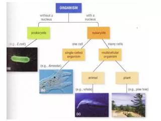
Prokaryotic Cells • Comparing prokaryotic and eukaryotic cells • Prokaryote comes from the Greek words for prenucleus. • Eukaryote comes from the Greek words for true nucleus.
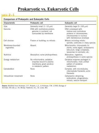
One circular chromosome, not in a membrane No histones No organelles Peptidoglycan cell walls Binary fission Paired chromosomes, in nuclear membrane Histones Organelles Polysaccharide cell walls Mitotic spindle Prokaryote Eukaryote
Average size: 0.2 -1.0 µm 2 - 8 µm • Basic shapes: Figures 4.1a, 4.2a, 4.2d, 4.4b, 4.4c
Unusual shapes • Star-shaped Stella • Square Haloarcula • Most bacteria are monomorphic • A few are pleomorphic Figure 4.5
Arrangements • Pairs: Diplococci, diplobacilli • Clusters: Staphylococci • Chains: Streptococci, streptobacilli Figures 4.1a, 4.1d, 4.2c
Glycocalyx • Outside cell wall • Usually sticky • A capsule is neatly organized • A slime layer is unorganized and loose • Extracellular polysaccharide allows cell to attach • Capsules prevent phagocytosis Figure 4.6a–b
Flagella • Outside cell wall • Made of chains of flagellin • Attached to a protein hook • Anchored to the wall and membrane by the basal body Figure 4.8a
Flagella Arrangement Figure 4.7
Motile Cells • Rotate flagella to run or tumble • Move toward or away from stimuli (taxis) • Flagella proteins are H antigens (e.g., E. coli O157:H7)
Motile Cells Figure 4.9
Motile Cells PLAY Animation: Bacterial Motility Figures 4.9a, 4.23d
Axial Filaments • Endoflagella • In spirochetes • Anchored at one end of a cell • Rotation causes cell to move Figure 4.10a
Fimbriae allow attachment • Pili are used to transfer DNA from one cell to another Figure 4.11
Cell Wall • Prevents osmotic lysis • Made of peptidoglycan (in bacteria) Figure 4.6a–b
Peptidoglycan • Polymer of disaccharideN-acetylglucosamine (NAG) and N-acetylmuramic acid (NAM) • Linked by polypeptides Figure 4.13a
Thick peptidoglycan Teichoic acids In acid-fast cells, contains mycolic acid Thin peptidoglycan No teichoic acids Outer membrane Gram-Positive Gram-Negative Cell Walls Cell Walls
Gram-Positive Cell Walls • Teichoic acids • Lipoteichoic acid links to plasma membrane • Wall teichoic acid links to peptidoglycan • May regulate movement of cations. • Polysaccharides provide antigenic variation. Figure 4.13b
Gram-Negative Outer Membrane • Lipopolysaccharides, lipoproteins, phospholipids • Forms the periplasm between the outer membrane and the plasma membrane. • Protection from phagocytes, complement, and antibiotics • O polysaccharide antigen, e.g., E. coli O157:H7 • Lipid A is an endotoxin • Porins (proteins) form channels through membrane.
Gram-Negative Outer Membrane Figure 4.13c
Gram Stain Mechanism • Crystal violet-iodine crystals form in cell. • Gram-positive • Alcohol dehydrates peptidoglycan • CV-I crystals do not leave • Gram-negative • Alcohol dissolves outer membrane and leaves holes in peptidoglycan. • CV-I washes out
Atypical Cell Walls • Mycoplasmas • Lack cell walls • Sterols in plasma membrane • Archaea • Wall-less or • Walls of pseudomurein (lack NAM and D amino acids)
Damage to Cell Walls • Lysozyme digests disaccharide in peptidoglycan. • Penicillin inhibits peptide bridges in peptidoglycan. • Protoplast is a wall-less cell. • Spheroplast is a wall-less Gram-positive cell. • L forms are wall-less cells that swell into irregular shapes. • Protoplasts and spheroplasts are susceptible to osmotic lysis.
Plasma Membrane Figure 4.14a
Plasma Membrane • Phospholipid bilayer • Peripheral proteins • Integral proteins • Transmembrane proteins Figure 4.14b
Fluid Mosaic Model • Membrane is as viscous as olive oil. • Proteins move to function. • Phospholipids rotate and move laterally. Figure 4.14b
Plasma Membrane • Selective permeability allows passage of some molecules • Enzymes for ATP production • Photosynthetic pigments on foldings called chromatophores or thylakoids
Plasma Membrane • Damage to the membrane by alcohols, quaternary ammonium (detergents), and polymyxin antibiotics causes leakage of cell contents.
Movement Across Membranes • Simple diffusion: Movement of a solute from an area of high concentration to an area of low concentration. • Facilitative diffusion: Solute combines with a transporter protein in the membrane.
Movement Across Membranes Figure 4.17
Movement Across Membranes • Osmosis: The movement of water across a selectively permeable membrane from an area of high water concentration to an area of lower water. • Osmotic pressure: The pressure needed to stop the movement of water across the membrane. Figure 4.18a
Movement Across Membranes Figure 4.18a–b
Movement Across Membranes • Active transport of substances requires a transporter protein and ATP. • Group translocation of substances requires a transporter protein and PEP. PLAY Animation: Membrane Transport
Cytoplasm • Cytoplasm is the substance inside the plasma membrane. Figure 4.6a–b
Nuclear Area • Nuclear area (nucleoid) Figure 4.6a–b
Ribosomes Figure 4.6a–b
Ribosomes Figure 4.19
Inclusions Figure 4.20
Metachromatic granules (volutin) Polysaccharide granules Lipid inclusions Sulfur granules Carboxysomes Gas vacuoles Magnetosomes Phosphate reserves Energy reserves Energy reserves Energy reserves Ribulose 1,5-diphosphate carboxylase for CO2 fixation Protein covered cylinders Iron oxide (destroys H2O2) Inclusions
Endospores • Resting cells • Resistant to desiccation, heat, chemicals • Bacillus, Clostridium • Sporulation: Endospore formation • Germination: Return to vegetative state
Eukaryotic Cells • Comparing prokaryotic and eukaryotic cells • Prokaryote comes from the Greek words for prenucleus. • Eukaryote comes from the Greek words for true nucleus.
Flagella and Cilia Figure 4.23a–b
Microtubules • Tubulin • Nine pairs + two arrangements Figure 4.23c
Cell Wall • Cell wall • Plants, algae, fungi • Carbohydrates • Cellulose, chitin, glucan, mannan • Glycocalyx • Carbohydrates extending from animal plasma membrane • Bonded to proteins and lipids in membrane
Plasma Membrane • Phospholipid bilayer • Peripheral proteins • Integral proteins • Transmembrane proteins • Sterols • Glycocalyx carbohydrates