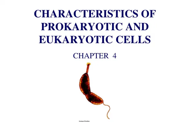
Prokaryotic and Eukaryotic Cells
150 likes | 913 Views
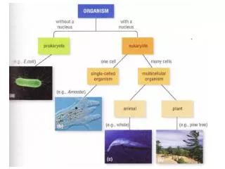
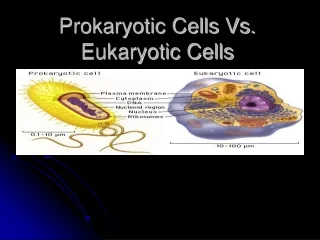
Prokaryotic and Eukaryotic Cells. A multimedia presentation to illustrate the differences and similarities between prokaryotic and eukaryotic cells and identify the role and function of the organelles within these cells. Click to get to Eukaryotic Cell.

Prokaryotic and Eukaryotic Cells
E N D
Presentation Transcript
Prokaryotic and Eukaryotic Cells A multimedia presentation to illustrate the differences and similarities between prokaryotic and eukaryotic cells and identify the role and function of the organelles within these cells.
Click to get to Eukaryotic Cell Click on each organelle to discover its role and function Prokaryotic Cell Structure
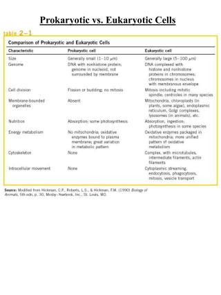
Click to get back to Prokaryotic Cell Prokaryotic Organelles Cytoplasm Nucleoid • The discovery of cytoplasm occurred in 1835 but unlike other cell organelles the credit of the discovery of cytoplasm cannot be attributed to a single scientist. • Cytoplasm is a thick, semi-transparent fluid found in both prokaryotic and eukaryotic cells that is detained by the plasma membrane of the cell. Cytoplasm occupies all the available space of the cell and allows all organelles to be suspended within it. Cytoplasm is the site where cell growth and expansion can occur and holds the cytoskeleton which creates movement and gives the cell its shape. Cytoplasmic streaming also allows the movement of various elements within the cell. Cytoplasm also has the role of being the site where cell reproduction, anaerobic glycolysis and cytokinesis can occur. The cytoplasm contains enzymes which break down macromolecules such as glucose into pyruvate to enable use by organelles, in this case, the mitochondria to produce and store energy. Cytosol, Organelles and Cytoplasmic inclusions comprise cytoplasm. • Cytosol is a gel-like substance that accounts for approximately 70% of the cell volume. Cytosol consists of the space of the cell not occupied by organelles. Inside cytosol can be found organic molecules, salt, water, enzymes, fatty acids, sugar and amino acids, ribosome's, proteasomes and soluble proteins. Protein filaments create the cytoskeleton which gives the cell its shape, these are found in the cytosol. • Cytoplasmic inclusions are insoluble substances such as starch, glycogen, crystals of some minerals and lipid droplets found in the cytoplasm. • In prokaryote cells the nucleoid region is the area where the nucleoid which is a long, single molecule of double stranded (helical), supercoiled DNA is found. The DNA of prokaryotes is referred to as a genophore which is compacted by super coiling. This DNA is different to eukaryotic cells which DNA consists of chromosomes compacted by chromatin found in a membrane enclosed organelle called the nucleus. The ends of the DNA genophore usually covalently bond together to for a physical and genetic circle. The DNA of the nucleoid is responsible for determining the production of proteins and enzymes within the cell and the chemical reactions which may occur. The reproduction of prokaryotic cells occurs through the asexual process of binary fission due to the nucleoid region not needing to divide. During binary fission the genophore is duplicated and attached to opposite sides of the cell membrane allowing the cell to then divide into two. The genophore then detaches forming the nucleoids of the two daughter cells. Free floating DNA called plasmids are also found in the nucleoid region.
Click to get back to Prokaryotic Cell Prokaryotic Organelles • Pili and fimbriae are both terms which describe short, thin, hair-like structures on the surface of prokaryotic cells. Pili, like flagella, are composed of proteins but are always shorter and stiffer than flagella, slightly smaller in diameter and usually numerous in quantity. Pili are most common in gram negative bacteria but can also occur in gram positive bacteria. Pili/fimbriae have the function of attaching to bacteria to surfaces, cells, tissues and substrates. This ability of pili to allow bacteria to attach to surfaces enables mass colonisation to occur and resistance against pathogens. Long conjugation pili also known as ‘F’ or sex pilus have the role of stabilizing mating bacteria during the function of reproduction. Flagella Pili • Flagella are long, obvious appendages that protrude from the cell body and usually have the general role of providing the cell with movement. Prokaryotic and Eukaryotic flagella are different through structure, protein composition and the mechanism of propulsion. The movement of the flagella is chemically driven. Some environmental nutrients attract motile prokaryotes while others repulse them. The reactive movement of the prokaryote is referred to as chemo taxis and involves the chemical gradient effecting the proton flow in the cell. Bacterial flagellum consist of three components which are the filament, hook and basal body. The filament of the bacterial flagellum is the rigid, helical, hollow structure which ascends from the cell surface. The filament is created from protein flagellin that is arranged in helical chains that detach from the ribosome's during synthesis to be transported through the hollow core and reattach to the filament in order to lengthen the flagellum. The hook of the flagellum connects the filament and basal body and is flexible. The basal body consists of rings and a rod that connect the flagellum to the cell wall and the cytoplasm membrane. The basal body enables the flagellum to rotate to create movement by acting as a rotary motor.
Click to get back to Prokaryotic Cell Prokaryotic Organelles Ribosomes Cytoplasmic Membrane • In every prokaryotic cell there are 60,000 ribosome's. Ribosome's found in prokaryotic cells are approximately 25nm in diameter and70S (S equals the density unit Svedberg) compared to 80S in eukaryotic cells. Prokaryotic ribosome's are separated into a smaller and a larger subunit. The larger subunit is 50S and the smaller subunit is 30S. Ribosome's can be found within the cell either free flowing in the cytosol (free ribosome's) where protein produced is excreted into the cytoplasm or attached to the endoplasmic reticulum (bound ribosome's) where the protein produced is usually transported to the cell membrane. The position of ribosome's within the cell is interchangeable based on the cell’s protein production needs. Ribosome's have the role of organising amino acids into specific proteins used for essential activities within the cell (protein synthesis). The function of ribosome's occurs where the DNA produces RNA or mRNA by the process DNA transcription which then the genetic code from the mRNA joins with the ribosome's to synthesise proteins. • Both prokaryotic and eukaryotic cells have a plasma membrane which is the semi-porous barrier that holds the shape of the cell, encloses all the organelles of the cell, protects the cell and is responsible for determining the organic and inorganic molecules that are able to pass through the plasma membrane (barrier) either into or out of the cell. The plasma membrane is semi-permeable which means that the barrier is selective to what is able to pass through the membrane. Small molecules such as oxygen, carbon and water have the ability to diffuse through the barrier, passing freely into or out of the cell. While, larger molecules such as glucose rely on the proteins embedded in the plasma membrane to pass though the barrier via active transport. The plasma membrane has a unique structure created by phospholipids known as the fluid mosaic model. The plasma membrane contains two phospholipids bilayers. The phosphate ends of the phospholipids are hydrophilic (water loving) and are directed towards the outsides of the bilayer while the lipid tail ends of the phospholipids are hydrophobic (water fearing) and are orientated towards the centre of the bilayer. The proteins embedded in this fluid mosaic model (the plasma membrane) have carbohydrates attached to their outer surfaces if they are partly exposed outside the membrane causing them to be referred to as glycoprotein's.
Click to get back to Prokaryotic Cell Prokaryotic Organelles Cell Wall Capsule • Prokaryotic cells have a rigid, tough and sometimes flexible cell wall located on the outside of the cytoplasmic membrane which provides the cell with protection from its environment, structural support an acts as a filtering system. The prokaryotic cell wall is composed peptidoglycan, a polymer created by interlocking chains of identical monomers. This peptidoglycan molecule is derived from two different types of glucose strands. Bacterial cell walls have two major variations in composition known as gram-positive or gram-negative cells. In gram-positive cells peptidoglycan creates approximately 90% of the cell wall while in gram-negative cells peptidoglycon composes only 5-20% of the cell wall but this barrier lies within the cytoplasmic membrane, it is not the outermost layer of the cell. • The capsule of prokaryotes is the outermost layer created with polysaccharides. A true prokaryotic layer is a discrete and detectable polysaccharide covering of the cell wall however less discrete layers known as slime layers or biofilm are often also referred to as capsules. Glycoalyx is a capsule created by a thin layer of polysaccharide fibres which occur on the surface of cells in nature. Capsules have a variety of functions including attaching to cell surfaces, protecting cells from bacterial engulfment of predatory protozoa, or phagocytes/white blood cell attack as antimicrobial agents. Capsules also function to protect the cells organelles from drying out and can become reserves of carbohydrates. Dental plaque is a form of biofilm which is created by oral bacteria.
Click to get to Prokaryotic Cell Click on each organelle to discover its role and function Eukaryotic Cell Structure
Click to get back to Eukaryotic Cell Eukaryotic Organelles Smooth and Rough Endoplasmic Reticulum • The endoplasmic reticulum in eukaryotic cells is an organelles that consists of a complex network of tubules and sacs called cisternae. Inside all these tubules and sacs which are connected by a single membrane of the endoplasmic reticulum is found a large, single, internal space called the cisternal space which is filled up with cytosol(intracellular fluid). The endoplasmic reticulum also has a perinuclear space which is the space between the inner and outer nuclear membranes that cover the nucleas of a cell. There are three different types of endoplasmic reticulum, smooth endoplasmic reticulum, rough endoplasmic reticulum and sacroplasmic reticulum. In most cells both smooth and rough endoplasmic reticulum is present. The function of the endoplasmic reticulum is based on the variety of endoplasmic reticulum. The function of smooth endoplasmic reticulum is to transport the protein molecules manufactured by the rough endoplasmic reticulum around the cell. The smooth endoplasmic reticulum is a small part of the endoplasmic reticulum but is prominant in cells found in teh liver, adrenal cortex and muscles. Smooth endoplasmic reticulum also has the role of carbohydrate metabolism, regulation of calcium ions, synthesis of steroids and lipids etc. The rough endoplasmic reticulum is called rough because it is studded with ribosomes that make the endopamic reticulum appear bumpy under microscope magnification. The ribosomes of teh rough endoplasmic reticulum is where proteins are made fromn the amino acids. The creation of particular proteins is caused by mRNA. This process allows proteins to be made to teh exact code neccessary with the help of tRNA.
Click to get back to Eukaryotic Cell Eukaryotic Organelles Nucleus and Nucleolus Nuclear Pores and Nuclear Envelope • Most eukaryotic cells contain a membrane bound, usually spherical, nuclease. Most eukaryotic cells only have one nuclease known as uninucleate while other cell can have two forming binucleate cells , or many forming multinucleate cells. In some cells, a nucleus isn’t present at all like in red blood cells, this is called enucleate. The nucleus is enveloped by a double membrane and approximately fills 15% of the total cells area. The nucleus holds a fine chromosome network which contains the strings of DNA molecules known as chromatin. The chromatin is classified into two groups heterochromatin which is an inactive form and euchromatin which is used in the process of transcribing the cell. The chromatin network inside the nucleus is in a fluid medium called nucleoplasm or also known as karyolymph. Inside the nucleus is a dense sphere known as the nucleolus. The nucleolus contains nucleotides that are involved in the process of making ribosome's made up of RNA and proteins which are transported to the cytoplasm and attached to the endoplasmic reticulum. The nucleus is responsible for storage of genes, chromosomes and RNA, selective transportation, production of ribosome's and production of mRNA. • The eukaryotic nuclear membrane also known as the nuclear envelope covers around the contents of the nucleus. The nuclear membrane function is to separate the nucleopasm from the cytoplasm and acts as a passable barrier for the exchange of micromolecules, ribonucleoproteins and ions. The nuclear envelope is made of two membranes called the inner and the outer membrane. Each membrane is formed with a lipoproteinous nature. The space between the two membranes is called the perinuclear cisternae. The outer membrane appears rough due to the attachment of ribosome's compared to the inner membrane which is a dense layer known as the ‘lamina densa’. The nuclear membrane can sometimes be continuous with the Golgi apparatus, mitochondria and endoplasmic reticulum. The nuclear membrane has thousands of nuclear pores. The nuclear pores act as an aqueous channel that connects the cytosol and the nucleoplasm. When materials such as ribosomal subunits are to pass through the pore opens up to approximately 25 nanometres wide creating the appropriate channel.
Click to get back to Eukaryotic Cell Eukaryotic Organelles Plasma Membrane Golgi Apparatus • Both prokaryotic and eukaryotic cells have a plasma membrane which is the semi-porous barrier that holds the shape of the cell, encloses all the organelles of the cell, protects the cell and is responsible for determining the organic and inorganic molecules that are able to pass through the plasma membrane (barrier) either into or out of the cell. The plasma membrane is semi-permeable which means that the barrier is selective to what is able to pass through the membrane. Small molecules such as oxygen, carbon and water have the ability to diffuse through the barrier, passing freely into or out of the cell. While, larger molecules such as glucose rely on the proteins embedded in the plasma membrane to pass though the barrier via active transport. The plasma membrane has a unique structure created by phospholipids known as the fluid mosaic model. The plasma membrane contains two phospholipids bilayers. The phosphate ends of the phospholipids are hydrophilic (water loving) and are directed towards the outsides of the bilayer while the lipid tail ends of the phospholipids are hydrophobic (water fearing) and are orientated towards the centre of the bilayer. The proteins embedded in this fluid mosaic model (the plasma membrane) have carbohydrates attached to their outer surfaces if they are partly exposed outside the membrane causing them to be referred to as glycoproteins. • The Golgi apparatus is an organelle that is found in all types of eukaryotic cells and is not connected to any of the other cell organelles nor is it attached to the cell membrane. The Golgi apparatus has the appearance of a stack of pancakes – these ‘pancakes’ are two layers of membrane surrounding a liquid filled centre known as cisternae and the number of cisternae in the ‘pancake stack’ depends on the variety of cell however there are usually five to eight cisternae per Golgi apparatus. Each Golgi apparatus has two sides the ‘cis’ face and the ‘trans’ face. The cis face of the Golgi body is located closest to the rough endoplasmic reticulum and nuclear membrane while the trans face is directed towards the plasma membrane of the cell. The main function of the Golgi apparatus is to process the synthesised proteins from the endoplasmic reticulum. This occurs when vesicles are sent from the endoplasmic reticulum attach to the cis face of the Golgi apparatus. The contents of the ER vesicle is released into the Golgi membrane and these molecules make there way through the cristenae and are modified by enzymes in the cisternae aided by carbohydrates and phosphates from nucleotides in the cytosol. Once the modifications have finished the molecule leaves the Golgi body through the trans face of the complex and is directed towards the membrane of the cell for exocytosis. The Golgi apparatus also creates lysosomes.
Click to get back to Eukaryotic Cell Eukaryotic Organelles Centrioles Lysosomes • Centrioles are structures found only within eukaryotic animal cells, plants cells and prokaryotic cells do not contain centrioles. The main role of the centriole is to produce microtubules as part of the cytoskeleton. The cytoskeleton prides the cell shape and motility. There are two centrioles within each animal cell which are found in the small region near the nucleus. These two centrioles are formed by 9 groups of 3 parallel microtubules which create a vague pinwheel shape. The centrioles are located perpendicular to one another inside the centrosome. Centrioles are 700 nanometres in length and 250 nanometres in diameter. The centrioles main function occurs during cell division. When cell division is to occur the centrioles duplicate creating to centrosomes with two centrioles each. The two centrosomes move to either end of the cell, opposite the nuclease, when thread like microtubules appear known as spindles (mitotic), the spindles are responsible for separating the single parent cell into two daughter cells with the replication and division of the chromosomes. Centrioles are also involved in the spatial arrangement of the organelles within the cell. • Lysosome s are membrane bound organelles found in the cytoplasm of eukaryotic cells. The word lysosome originates from two Greek words ‘lysis’ which means destruction and ‘soma’ which means body. Lysosomes were discovered in 1949 by cytologist Christian de Duve from Belgian. Lysosomes usually have a spherical shape - they share similarities in structure to vacuoles and have a diameter of approximately 0.25 µm. Lysosomes contain 50 types of hydrolytic enzymes and an acidity of about 4.8 pH which without the lysosome membrane would destroy the rest of the cell. Lysosomes are formed by the Golgi apparatus as ‘budded’ membrane. The main function of lysosomes is to break down all the cellular waste products, fats, carbohydrates and proteins into simple compounds which can been then returned to the cytoplasm as new cell building materials. Lysosomes are also known as ‘suicidal sacs or bags’ because they can digest the entire cell. Other functions of lysosomes is destruction of worn out cell parts, destruction of old red blood cells and dead cells, defence against invading bacteria, dissolving blood clot.
Click to get back to Eukaryotic Cell Eukaryotic Organelles Microfilaments and Microtubules Mitochondria • There are three types of filaments that make up the cytoskeleton. These are the microfilaments, microtubules and intermediate filaments. The cytoskeleton is responsible for cell shape, movement (motility) and the motility of the organelles within the cell. Microfilaments, microtubules and intermediate filaments all are created by proteins and have the ability to self-assemble into a filamentous network where needed within the cell. The three features of the cytoskeleton are very dynamic with the ability to lengthen and shorten rapidly which aids movement, cell division and migration. The role of the microfilament system is to hold important plasma membrane proteins, produce cell movement and aid cell division. The filaments of the microfilament system are approximately 6 nanometres in diameter and are composed of a protein called ‘actin’. The protein actin is found in to main states inside cells which are G-actin (for globular actin) and F-actin (for filamentous actin). The difference between ‘G’ and ‘F’ actin is the concentration of actin protein. Microfilaments are polarised with a fast growing ‘plus’ end and a slow growing ‘minus’ end. The fast growing ‘plus’ ends of the microfilaments are generally orientated towards the edge of the cell. Microtubules share the same polarised features and consist of proteins like microfilaments however microtubules are situated close to the nucleus not the edge of the cell and have a diameter 25 nanometres in length. The main function of microtubules is to act as a transport system for vesicles for exocytosis. • Mitochondria are responsible for converting the energy provided from the food we consume, carbohydrates, lipids and proteins into useable energy in the form of ATP (adenosine triphosphate). This conversion of energy within the mitochondria is known as aerobic respiration (oxidative phosphorlation). This causes the mitochondria to be sometimes known as the ‘powerhouses’ of the cell. Mitochondria also have involvement in the processes of cell division and cell death. Mitochondria are generally rod shaped organelles which are approximately 1-10 micrometers in length. Mitochondria are quite flexible with the ability to change shape quickly and move throughout the cell constantly. The concentration of mitochondria within a cell is dependent on the metabolic activity levels within that cell. Muscle cells have high concentrations of mitochondria due to having large metabolic activity in working muscles. Mitochondria have two membranes, the outer and inner membranes. Both membranes are made of a phospholipids bilayer and proteins. The outer membrane of the mitochoondria is smooth and is permeable to nutrient molecules, ions, ATP and ADP molecules. The inner membrane of the mitochondria contains an electron transport chain and is permeable only to oxygen, carbon dioxide and water. The inner membrane folding are known as cristae and these cristae increase the surface area where ATP can be produced. The two membranes divide the intermembrane space which is between the inner membrane and the outer membrane and the mitochondrial matrix found enclosed by the inner membrane. Cellular respiration occurs in the mitochondrial matrix due to the high concentration of enzymes.
Click to get back to Eukaryotic Cell Click to get back to Prokaryotic Cell Bibliography • http://www.buzzle.com/articles/structure-and-functions-of-cytoplasm.html • http://www.iscid.org/encyclopedia/Nucleoid • http://student.ccbcmd.edu/courses/bio141/lecguide/unit1/prostruct/nucleoid.html • http://en.wikipedia.org/wiki/Nucleoid • http://www.suite101.com/content/external-structures-of-prokaryotic-cells-a100536 • http://science.jrank.org/pages/2725/Flagella.html • http://en.wikipedia.org/wiki/Flagellum • http://student.ccbcmd.edu/courses/bio141/lecguide/unit1/prostruct/flag.html • http://biology.about.com/od/cellanatomy/p/ribosomes.htm • http://www.tutorvista.com/biology/function-of-ribosome-cell • http://www.buzzle.com/articles/ribosomes-function.html • http://www.tutorvista.com/biology/prokaryotic-cell-ribosome • http://www.suite101.com/content/ribosome-structure-and-function-principles-facts-a156745 • http://student.ccbcmd.edu/courses/bio141/lecguide/unit1/prostruct/proribo.html • http://micro.magnet.fsu.edu/cells/plasmamembrane/plasmamembrane.html • http://www.bae.uky.edu/~snokes/bae549thermo/microbio/cellcharac.htm • http://www.suite101.com/content/prokaryotic-cells-a32315 • http://www.cellsalive.com/cells/bactcell.htm • http://student.ccbcmd.edu/courses/bio141/lecguide/unit1/prostruct/cm.html • http://www.suite101.com/content/bacterial-cell-wall-a32297 • http://wiki.answers.com/Q/Charisteristics_of_a_prokaryotic_cell_wall • http://www.textbookofbacteriology.net/structure_3.html • http://www.biologyreference.com/Co-Dn/Cytoskeleton.html • http://biology.about.com/od/cellanatomy/ss/mitochondria.htm • http://www.ruf.rice.edu/~bioslabs/studies/mitochondria/mitotheory.html • http://www.buzzle.com/articles/mitochondria-structure-and-functions.html • http://biology.about.com/gi/o.htm?zi=1/XJ&zTi=1&sdn=biology&cdn=education&tm=142&gps=504_118_1596_692&f=21&su=p897.10.336.ip_&tt=29&bt=0&bts=0&zu=http%3A//micro.magnet.fsu.edu/cells/mitochondria/mitochondria.html • http://www.buzzle.com/articles/centriole-function.html • http://www.buzzle.com/articles/centrioles.html • http://www.helium.com/items/1818212-role-of-centrioles-in-animal-cells • http://www.tutorvista.com/biology/lysosome-structure-and-function • http://lic.leidenuniv.nl/files/lic/images/nucleoid.jpg • http://www.buzzle.com/articles/lysosome-structure.html • http://www.daviddarling.info/encyclopedia/L/lysosome.html • http://www.brighthub.com/science/genetics/articles/22922.aspx • http://www.suite101.com/content/the-golgi-apparatus-a125802 • http://www.buzzle.com/articles/golgi-apparatus-function.html • http://www.spiritus-temporis.com/golgi-apparatus/function.html • http://www.buzzle.com/articles/cell-nucleus-structure-and-functions.html • http://www.buzzle.com/articles/nucleus-function.html • http://www.buzzle.com/articles/nucleolus-function.html • http://users.rcn.com/jkimball.ma.ultranet/BiologyPages/N/Nucleus.html#Nuclear_Pore_Complexes_(NPCs) • http://www.buzzle.com/articles/nuclear-membrane-function.html • http://www.tutorvista.com/biology/animal-nucleus • http://www.buzzle.com/articles/smooth-endoplasmic-reticulum.html • http://www.buzzle.com/articles/rough-endoplasmic-reticulum-function.html