Download
1 / 1
10 likes | 237 Views
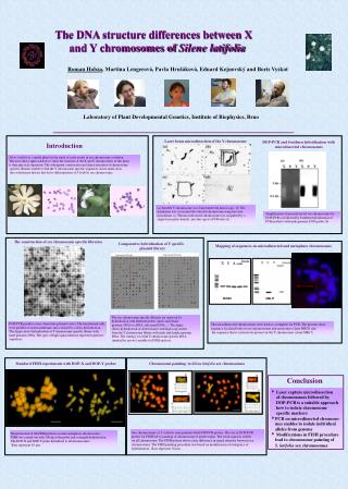
The DNA structure differences between X and Y chromosomes of Silene latifolia. Roman Hobza , Martina Lengerová, Pavla Hrušáková, Eduard Kejnovský and Boris Vyskot. Laboratory of Plant Developmental Genetics, Institute of Biophysics, Brno. Laser beam microdissection of the Y chromosome.

E N D
The DNA structure differences between X and Y chromosomes of Silene latifolia Roman Hobza, Martina Lengerová, Pavla Hrušáková, Eduard Kejnovský and Boris Vyskot Laboratory of Plant Developmental Genetics, Institute of Biophysics, Brno Laser beam microdissection of the Y chromosome DOP-PCR and Southern hybridization with microdissected chromosomes Introduction Silene latifolia is a model plant for the study of early events in sex chromosome evolution. The most direct approach how to study the structure of the X and Y chromosomes of this plant is their physical separation. The subsequent construction and characterisation of chromosome specific libraries enable to find the Y chromosome specific sequences and to make clear the evolutionary history that led to differentiation of S.latifolia sex chromosomes. male female (a) Suitable Y chromosome was found under the microscope. (b) The membrane was cut around the selected chromosome using the laser microbeam. (c) The microdissected chromosome was catapulted by a single laser pulse directly into the cap of a PCR tube (d). Amplification of microdissected sex chromosomes by DOP-PCR (a) followed by Southern hybridisation of PCR products with male genomic DNA probe (b). The construction of sex chromosome specific libraries Comparative hybridisation of Y specific plasmid library Mapping of sequences on microdissected and metaphase chromosomes MK17 probe RH20 MK17 The sex chromosome-specific libraries are analysed by hybridisation with different probes (male and female genomic DNA or cDNA, autosomal DNA...). The figure shows hybridisation of dotted clones with high copy inserts from the Y chromosome library with male and female genomic DNA. The strategy is to find Y chromosome specific DNA (marked by arrows) suitable for FISH analysis. DOP-PCR products were cloned into plasmid vector. The transformed cells were gridded on nylon membrane and screened by colony hybridisation. The figure shows hybridisation of Y chromosome specific library with male genomic DNA. The spots of high signal intensity represent repetitive sequences. The microdissected chromosomes were used as a template for PCR. The pictures show sequence localised both on sex chromosomes and autosomes (clone RH20) and the sequence that is exclusively present on the Y chromosome (clone MK17). Standard FISH experiments with DOP-X and DOP-Y probes Chromosome painting in Silene latifolia sex chromosomes Conclusion Laser capture microdissection of chromosomes followed by DOP-PCR is a suitable approach how to isolate chromosome specific markers PCR on microdissected chromoso- mes enables to isolate individual alleles from genome Modifications in FISH procedure lead to chromosome painting of S. latifolia sex chromosomes Sex chromosomes of S. latifolia were painted with DOP-PCR probes. The use of DOP-PCR probes for FISH led to painting of chromosome of probe origin. The weak signal is visible on all chromosome. The FISH pattern shows clear difference in signal intensity between sex chromosomes. The FISH painting procedure was based on modification of stringency of hybridisation . Bars represent 10 µm. Demonstration of the FISH patterns on male metaphase chromosomes. FISH was carried out with 100 ng of the probe and overnight hybridisation. The DOP-X and DOP-Y probe hybridised to all chromosomes. Bars represent 10 µm.