Download
1 / 64
680 likes | 1k Views
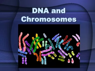
DNA and Chromosomes. Genes are carried by Chromosomes Two plant cells visualized by light microscope, DNA stained with DAPI. Chromosome in Cells DNA (deoxyribonucleic acid) AGTC Human 46 chromosomes 22 homologs, x, or x/y.

E N D
Genes are carried by Chromosomes Two plant cells visualized by light microscope, DNA stained with DAPI Chromosome in Cells DNA (deoxyribonucleic acid) AGTC Human 46 chromosomes 22 homologs, x, or x/y
Experimental procedures demonstrating that DNA is the genetic material 1940s
The Structure and Function of DNA • Genetic information is carried in the linear sequence of nucleotides in DNA • Genetic information contains instructions to synthesize proteins • DNA forms double helix with two complimentary strands holding together by hydrogen bonds between A-T (2 bonds) and G-C (3 bonds) • DNA duplication occurs using one strand of parental DNA as template to form complimentary pairs with a new DNA strand. • DNA is in nucleus in eucaryotes
1953 Watson and Crick determined the structure of DNA DNA and its Building Nucleotides: Guanine (G), Adenine (A), Cytosine (C), Thymine (T). Polarized strand, 5’->3’ Base inside, sugar outside
DNA and its Building Antiparallel strands
DNA Pairs A always pairs with T, and G with C, A-T two hydrogen bonds, G-C three hydrogen bonds
DNA Double Helix 10.4 nucleutides/turn; 3.4 nm between nucleutides
DNA to Protein Genome: the complete set of information in an organism’s DNA Total length of DNA about 2 meters long in a human cell, encoding about 30000 proteins
To carry the genomic information to daughter cells DNA Duplication Using itself as template
Cell Nucleus, compartmentalized DNA activity Nuclear pores allow communication Nuclear lamina and cytoskeleton mechanically support the nucleus
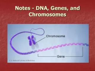
Chromosomal DNA and its Packaging • A gene is a nucleotide sequene in a DNA molecule that act as a functional unit for protein production, RNA synthesis. • Introns and Exons • Chromosome: single long DNA contains a linear array of many genes. • Human genome contains 2.3x10⁹ DNA nucleotide pairs, with 22 different autosomes and 2 sex chromosomes. • Chromosomal DNA: replication origins, telomeres, centromeres • Histones form the protein core for DNA wrapping • Nucleosome: repeating array of DNA-protein particles • Modification of Chromatin and nucleosomes: histone H1, ATP-driven chromatin remodeling complexes, and enzymatically catalyzed covalent modification of the N-terminal tails of Histones
Human Chromosome Complex of DNA and protein is called chromatin 44 homologous chromosomes and 2 sex chromosomes Complementary DNA with different Dyes The arrangement of the full chromosome set is called karyotype
Banding Pattern of human chromosomes Giemsa Staining Green line regions: centromeres Encoding ribosome
Conservation between human and mouse genomes Usually important genes are encoded by conserved regions Note: Human chromosome 1 and mouse chromosome 4 mouse human centromere
Cell Cycle DNA molecule not only carries genetic information, but also undergoes conformational change Chromosomes exist through the cycle Mitotic and interphase chromosome Single chromosome can only be visible during mitosis
Three important DNA sequences Telomere, replication origin, centromere
DNA Molecules are highly condensed in chromosomes Nucleosomes of interphase under electron microscope Nucleosome: basic level of chromosome/chromatin organization Chromatin: protein-DNA complex Histone: DNA binding protein A: diameter 30 nm; B: further unfolding, beads on a string conformation
Nucleosome Structures Histone octamer 2 H2A 2 H2B 2 H3 2 H4
X-ray diffraction analyses of crystals Structure of a nucleosome core particle
The bending of DNA in a nucleosome 1. Flexibility of DNAs: A-T riched minor groove inside and G-C riched groove outside 2. DNA bound protein can also help
Irregularities in the 30-nm fiber Flexible linker, DNA binding proteins Structural modulators: H1 histone, ATP-driven Chromatin remodeling machine, covalent modification of histone tails
Covalent Modification of core histone tails Acetylation of lysines Mythylation of lysines Phosphorylation of serines Histone acetyl transferase (HAT) Histone deacetylase (HDAC)
Summary • DNA, Chromosome • Centromere, telomere, replication origin • Nucleosome, Chromatin, • Histone: H1, H2A, H2B, H3, H4 • Histone octamer, DNA packaging • DNA binding proteins, Histone modifications
The Global Structure of Chromosomes • Some rare cases of interphase chromosomes, certain features maybe universal • Representative forms forming typical interphase chromosome • Chromosome at mitosis
A model for the structure of a lampbrush chromosome Chromomeres: highly condensed and in general not expressed until unfolding
A polytene chromosome from Drosophila salivary gland Dark bands and interbands
Chromosome puffs Folding and refolding at a time course of 22 hours
RNA synthesis in Chromosome puffs Red: newly synthesized BrUTP; Blue: old ones diffused
Position Effects on Gene Expression Heterochromatin: condensed Euchromatin: loose
Speculative Model for the heterochromatin at the ends of yeast chromosomes Sir: Silent information regulator binding to unacetylated histone tails
Speculative Model for the heterochromatin at the ends of yeast chromosomes DNA-binding proteins recognize DNA sequence close to telomere, recruit Sir proteins and cause histone tail modification, forming heterochromatin