Download
1 / 37
1.78k likes | 6.29k Views
Ascending Tracts of the Spinal cord. Dr Rania Gabr. Objectives. Define the meaning of a tract. Distinguish between the different types of tracts. Locate the position of each tract. Describe the sensory pathway.

E N D
Ascending Tracts of the Spinal cord Dr Rania Gabr
Objectives • Define the meaning of a tract. • Distinguish between the different types of tracts. • Locate the position of each tract. • Describe the sensory pathway. • Describe gracile and cuneate tracts and pathways for conscious proprioception, touch, pressure and vibration from the limbs and trunk. Describe lateral spinothalamic tract and pathways for pain and temperature from the limbs and trunk. • Describe ventral spinothalamic tract and pathways for simple touch from the limbs and trunk. • Describe dorsal and ventral spinocerebellar tracts and pathways for unconscious proprioception from the limbs and trunk. • Describe Reticulothalamic and spinothalamic tracts for dull aching pain
TRACTS • Bundles or fasciculi of fibers that occupy more or less definite positions in the white matter. • They have the same Origin, Termination and carry the same Function. • Theyserve to join the brain to the spinal cord. • They are classified into: 1-Ascending (sensory or afferent). 2-Descending (motor or efferent).
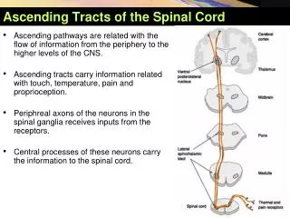
Ascending Tracts • Carry impulses from pain, thermal, tactile, muscle and joint receptors to the brain. • Some of this information eventually reaches a conscious level (the cerebral cortex), • while some is destined for subconscious centers (e.g. the cerebellum). • Ascending sensory pathways between the peripheral receptors and the cerebral cortex, are organized in a three neuronal chain: - First order neurone- Second order neurone- Third order neurone
The first-order neuroneor (primary afferent neurone) : • enters the spinal cord through the dorsal root of a spinal nerve and its cell body lies in the dorsal root ganglion.
The main fibers remain on the ipsilateral side of the cord and terminate by syapses with the second neuroneeither in the spinal grey matter or in the medulla oblongata of the brain stem.
The second order neurone • Has its cell bodies in thecordor medulla oblongata. • Its axon cross over (decussates) to the opposite side of the CNS and ascend to the thalamus, where it terminate upon the third neurone. • The third-order neuroneHas its cell bodies in the thalamus. • Its axons pass to the somatosensory cortex of the parietal lobe of the cerebral hemisphere.
VPL SECOND ORDER NEURON 2nd 1st • cross the mid line • in front of central canal
Ascending Tracts • Dorsal column tracts • Spinothalamic tracts( Ventral AND Lateral) • Posterior spinocerebellar • Anterior spinocerebellar • Cuneocerebellar • Spinotectal • Spinoreticular • Spino-olivary
Functions of Ascending Tracts • Lateral spinothalamic • Anterior spinothalamic • Posterior column • Anterior and posterior spinocerebellar and cuneocerebellar tracts • Spinotectal • Spinoreticular • Painful and thermal sensations • Crude touch and pressure • Descriminative fine touch and joint and muscle information and vibration sense • Muscle and joint unconscious information • Pain, thermal and tactile information to superior colliculus • Muscle and joint information to reticular formation
Ascending Or Sensory Tracts 1- Dorsal column tracts: (Gracile & Cuneate) Function:Transmit • Proprioceptive(deep) sensations (sense of movement, position, vibration). • Fine touchsensations (tactile localization, tactile discrimination & stereognosis). These senses reach a Consciouslevel (cerebral cortex).
2- Spinothalmic tracts: Function: Transmit impulses concerned with specific sensory modalities: pain, temperature, touch, that reach a Consciouslevel (cerebral cortex). 3- Spinocerebellar tracts: Function: Carries unconsciousinformation from muscles, joints, skin, subcutaneous tissues Transmit impulses from tactile and stretch receptors to Subconsciouscenters (cerebellum) for muscle tone and coordinations.
1. Dorsal column TractsFasciculus gracilis and fasciculus cuneatus • Function: carry discriminative(fine) touch, proprioception: vibratory sense and conscious muscle joint sense • Inputs from pacinian corpuscles, Messiner’s corpuscles, joint receptors, muscle spindles and Golgi tendon organs ) Axon of 1st order neuron enter the spinal cord through the dorsal roots of the spinal nerves. • passes directly to the posterior white column of the same side ( without synapsing )
Long ascending fibers travel upward in the posterior column of the same side as fasciculus gracilis and fasciculus cuneatus • Fibers of Fasciculus Gracilisenter via the sacral, lumbar and lower thoracic 6 levels; (lower limbs). • Fibers of Fasciculus Cuneatusenter via the upper 6 thoracic and cervical levels; (upper limbs). • Synapse on the 2nd order neuron in the nucleus gracilis and cuneatus of medulla oblongata of the same side.
[ nucleus G & C ] in medulla fasciculus cuneatus cervical segments upper 6 thoracic segments lower 6 thoracic segments lumbar segments sacral segments G C fasciculus gracilis
Axons of second-order neurones : • decussate in the medulla as the internal arcuate fibers(sensory decussation). • Then it ascendin (opposite side) through brain stem as the Medial Lemniscus. • The medial lemniscus ASCENDS through medulla oblongata, pons, and midbrain terminates in the ventral posterior (VP) nucleus of the thalamus. Prof. Makarem
Third-order neurone: • (thalamocortical, or sensory radiations- run in the internal capsule,coronaradiata to reach the postcentralgyrus of cerebral cortex area 3, 1 and 2 ) : somatosensory cortex.
Clinical application destruction of fasciculus gracilis and cuneatus • loss of muscle joint sense, position sense, vibration sense and tactile discrimination • on the same side • below the level of the lesion (extremely rare to have a lesion of the spinal cord to be localized as to affect one sensory tract only )
Spinothalamic tracts lie lateral and ventral to the ventral horn of the spinal grey matter. 2- Spinothalamic tracts • They carry painand thermal(temperature) sensationsand also non-discriminative (crude) touch and pressure. • The Lateral spinothalamic tracts carry pain and temperaturesensations. • The ventral spinothalamic tracts carry crude touch and pressure. • But fibers carrying these modalities are probably intermingled, at least to some extent. Prof. Makarem
A-Lateral Spinothalamic Tract (pain & temperature) • Function: • Carries pain & Temperature to thalamus and sensory area of the cerebral cortex. • Neurones: 3 Neurones • Neurone I: Small cells in the dorsal root ganglia. • Neurone II:Cells of substantia gelatinosa of Rolandi in the posterior horn. • Neurone III: Cells of (VPL) nucleus of the thalamus. • The spinothalamic tract contains second-order neurones, the cell bodies of which lie in the contralateral dorsal horn.
Pain and thermal impulses ( input from free nerve endings, thermal receptors ) • transmitted to spinal cord in delta A and C fibres • central process enters the spinal cord through Dorsal nerve root, • the central process of 1st order neuron synapse with cell body of 2nd order neuron in substantiagelatinosa of posterior gray column of the spinal cord
the axon of 2nd order neuron cross to the opposite side in the anterior white commissure and ascend in contralateralwhite column as lateral spinothalamic tract • end by synapsing with 3rd order neuron in the ventral posterolateral nucleus (VPL )of thalamus • axon of the 3rd order neuron passes through the posterior limb of internal capsule and corona radiata to reach the postcentralgyrus of cerebral cortex ( area 3, 1 and 2 )
Clinical application destruction of LSTT • loss of • pain and thermal sensation • on the contralateral side • below the level of the lesion patient will not respond to pinprick recognize hot and cold • Lesion: • Syringomyelia, (widening of the central canal) leads to Loss of pain & temperature below the level of the lesion.
B- Ventral Spinothalamic(Crude touch & Pressure) • Function: • Carries crude touch & pressure to thalamus and sensory cortex. • input from free nerve endings, Merkel’s tactile disks
Neurones: 3 Neurones • NeuroneI: Medium sized cells in the dorsal root ganglia. • Neurone II: Cells of main sensory nucleus or (nucleus proprius). cross to the opposite side (decussate) • ascend in the contralateral ventral column as ASTT • Ends in the VPL nucleus of the thalamus. • Neurone III: Cells of VPL nucleus of thalamus project to cerebral cortex ( area 3, 1 and 2 ) • Effect of lesion: Loss of crude touch sensation below the level of the lesion. on the contralateral side of the body
Axons carrying pain and temperaturedecussate within one segment of their origin, • while those carrying touch and pressuremay ascend for several segments before crossing.
It is highly organisedsomatotopically;consequently, the origin of sensory stimuli can be accurately localised. • It is thought to be the route via which sharp, pricking pain(sometimes called 'fast' pain) is conducted
3- Spinocerebellartracts • Ascending pathways that carry impulses to a subconscious level are represented by the spinocerebellar tracts. • Fibres of spinocerebellar tracts form dorsal and ventral tracts that are located at the dorsolateral & ventrolateral surfaces of the cord, respectively. Prof. Makarem
Both tracts carry information derived from muscle spindles, Golgi tendon organs and tactile receptorsto the cerebellum for the control of posture & coordination of movement.
The spinocerebellar system consists of a sequence of only two neurones. • Neurone I:Large cells of dorsal root ganglia. • Neurone II: cells of the nucleus dorsalisClark's nucleus. • give rise to axons ascending to the cerebellum of the same side • Both spinocerebellar tracts contain second-order neurones. • The tract neurons terminate directly in the cerebellar cortex. MOSTLY VERMIS Prof. Makarem
Dorsal (Direct) Spinocerebellar tract • Fibers of the dorsal spinocerebellar tractoriginate from the cells of Clarke's columnat the base of the posterior horn. • The axons ascend ipsilaterally to enter the cerebellum through the inferior cerebellar peduncle.
Ventral (Indirect) Spinocerebellar Tract • Fibers of the ventral spinocerebellar tractdecussate, ascend on the contralateral side of the cord and enter the cerebellum via the Superior cerebellar peduncle. • Some axons then recross within the cerebellar white matter.
Spinoreticulothalamic • The spinoreticulothalamic system represents an additional route by which sensory impulses ascend to higher centers. • Second-order neurons arising from the dorsal horn ascend in the ventrolateral region of the cord • They terminate in the brain stemreticular formation, particularly within the medulla. • Reticulothalamicfibres then ascend to the intralaminar thalamic nuclei, which in turn activate the cerebral cortex.
The spinoreticulothalamic system is thought to be the route via which dull, aching pain (sometimes called 'slow' pain) is transmitted to a conscious level. • Activation of spinothalamic and spinoreticular fibers, which may ultimately be perceived as unpleasant or painful, can be modulated by descending pathways from the brain.