Download
1 / 33
390 likes | 1.36k Views
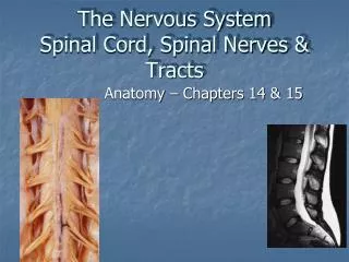
The Nervous System Spinal Cord, Spinal Nerves & Tracts. Anatomy – Chapters 14 & 15. The Spinal Cord. Begins at foramen magnum, runs through vertebral foramen (spinal canal), & ends at L2 vertebral level by forming conus medularis.

E N D
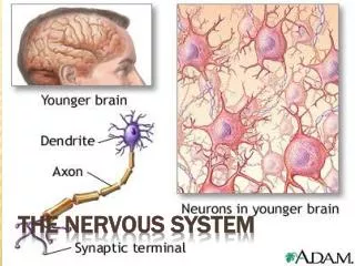
The Nervous SystemSpinal Cord, Spinal Nerves & Tracts Anatomy – Chapters 14 & 15
The Spinal Cord • Begins at foramen magnum, runs through vertebral foramen (spinal canal), & ends at L2 vertebral level by forming conus medularis • The spinal cord (as well as the brain) is well protected by bones, CT membranes (meninges), and fluid (cerebrospinal fluid (CSF))
Meninges Meninges– membranes that surround and protect the CNS • Three layers: • Dura mater • Arachnoid mater • Pia mater
Dura Mater – tough, fibrous CT outer membrane; one layer thick around spinal cord with epidural space external Arachnoid mater– “spidery” web-like middle layer Pia Mater – delicate, thin inner layer
Filum terminale - extension of pia mater extends from tip of cord to coccyx to anchor cord in place Denticulate ligaments - anchor cord laterally
Lumbar cystern Subarachnoid space – between arachnoid & pia mater; contains cerebrospinal fluid (CSF) Lumbar cistern – area of subarachnoid space below the conus medularis; site for lumbar puncture (“spinal tap”)
Spinal Cord Anatomy • Begins at foramen magnum & ends at L2 vertebral level by forming conus medularis • Has 2 thickened areas- cervical enlargement - supplies nerves to upper extremity • lumbar enlargement - supplies nerves to lower extremity • Made up of 31 spinal cord segments
Dorsal root ganglion (DRG) Dorsal root Ventral root • Each spinal cord segment has a pair of • dorsal roots with their associated dorsal root ganglia (DRG) • ventral roots
Each dorsal root contains the axons of sensory neurons • Each dorsal root ganglion contains the cell bodies of these sensory neurons • Each ventral root contains the axons of motor neurons
The dorsal & ventral roots of each segment come together at the intervertebral foramen (IVF) to form a mixed spinal nerve
Spinal Nerves • Part of the PNS • Contain both motor & sensory fibers • 31 pair of nerves – each nerve forms from union of dorsal/ventral root of spinal cord segment & exits between vertebra at IVF • 8 pair cervical spinal nerves – 1st cervical nerve exits between occipital bone & C1, 8th cervical nerve exits the IVF between C7-T1 • 12 pair thoracic spinal nerves • 5 pair lumbar nerves • 5 pair sacral nerves • 1 pair coccygeal nerves
Below the conus medularis, spinal nerves must angle downward (in the subarachnoid space) before exiting their IVF/sacral foramina. These spinal nerves make up the cauda equina Cauda equina
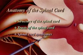
Posterior median sulcus Posterior column Posterior gray horn - sensory Central canal Lateral column Lateral gray horn (T1-L2, S2-S4) - autonomic Anterior gray horn - motor Anterior column Anterior median fissure Sectional Anatomy of the Spinal cord Gray commissure
The spinal cord has a narrow central canal surrounded by “horns” of gray matter connected by commissures. Gray matter horns contain sensory & motor nuclei (groups of cell bodies) & glial cells. Gray matter is surrounded by white matter “columns” (aka funiculi) which are made up of groups of myelinated axons creating organized ascending & descending tracts (aka fasciculi).
Tracts (Motor & Sensory Pathways)(Chap. 15) • Groups of axons found in the white matter columns of the spinal cord that carry specific information • Ascending tracts - carry sensory information up the spinal cord to areas of the brain (eventually terminating in cerebrum or cerebellum) • Descending tracts – carry motor information from the brain down to specific levels of the spinal cord (eventually terminating on skeletal muscles)
Ascending Tracts • Three major groups of pathways transmit somatic sensory information originating from receptors, up the spinal cord to the brain – • Spinothalamic tracts • Posterior column pathways • Spinocerebellar tracts
Spinothalamic tracts Anterior spinothalamic tract (ASTT) – crude touch & pressure Lateral spinothalamic tract (LSTT) – pain & temperature THALAMUS
Posterior Column Pathways • Fasciculus cuneatus & fasciculus gracilis – • “conscious” proprioception (joint position) • discriminitive (fine) touch (2-point discrimination, stereognosis, graphism) • vibration • pressure
Spinocerebellar Tracts Anterior spinocerebellar tract (ASCT) & Posterior spinocerebellar tract (PSCT) – • “unconscious” proprioception (from golgi tendon organs, muscle spindles & joint capsules) • muscle tone • balance
Descending Pathways • Carry motor signals from conscious & unconscious areas of the brain, down the spinal cord to control contraction of skeletal muscles • Corticospinal pathways (aka Pyramidal tracts)- include anterior & lateral corticospinal tracts, and corticobulbar tracts) – conscious motor control • Subconscious Motor Pathways – include medial and lateral pathways
Corticospinal (Pyramidal) Pathways • Corticobulbar tracts – voluntary control of skeletal muscles of head & neck through cranial nerves • Lateral corticospinal tracts (LCST) – voluntary control of skeletal muscles in neck & body; fibers cross in pyramidal decussation of M.O. • Anterior corticospinal tracts (ACST) - voluntary control of skeletal muscles in neck & body; fibers cross at spinal cord level in anterior commissure
Medial & Lateral Pathways Originate from subconscious areas of the brain and are integrated with corticospinal pathways to allow for coordination of motor activity, maintenance of posture and muscle tone Medial pathways – unconscious control over head, eyes, neck, trunk & proximal limb muscles for gross muscle movements; include vestibulospinal, tectospinal, & reticulospinal tracts Lateral pathways – unconscious control over distal limb muscles for precise muscle movements; include rubrospinal tracts
In order for sensory information to enter the spinal cord and ascend in a sensory tract, and for motor information to get from a descending tract to reach a skeletal muscle, impulses must travel through peripheral nerves (spinal nerves & cranial nerves)
Spinal Nerves • 31 pair • Part of PNS • Formed by union of ventral (motor) root and dorsal (sensory) root
Once formed, spinal nerves will branch into Rami • Dorsal ramus – transmits sensations from skin of back & neck; provides motor control of deep muscles of back; found at all spinal nerves
Ventral ramus – provides motor control to muscles of extremities, anterior & lateral trunk; transmits sensations from all but skin of back; found at all spinal nerves
Rami communicantes(white ramus & gray ramus) – carry autonomic motor fibers (ANS) to smooth muscles & glands in ventral body cavity; transmit visceral sensations; only found at T1-L2 spinal nerves
Nerve Plexuses Adjacent ventral rami will form complex interwoven networks of nerve fibers (axons) known as a nerve plexus Four plexuses – cervical, brachial, lumbar, & sacral Emerging from each plexus will be specifically named peripheral nerves, which will contain fibers from multiple spinal cord levels
Cervical plexus (C1-C5) Motor control for muscles of neck region, levator scapulae, scalenes, SCM & trapezius (along with CN XI), and diaphragm Sensory from upper chest, shoulder, neck & ear regions • Phrenic nerve (C3-C5)
Brachial plexus (C5-T1) Motor to & sensory from pectoral girdle region & upper extremities • Axillary nerve (C5-C6) • Musculocutaneous nerve (C5-7) • Radial nerve (C5-T1) • Median nerve (C6-T1) • Ulnar nerve (C8-T1)
Lumbar plexus (T12-L4) Motor to muscles of abdominal, pelvic and lower extremity (anterior & medial) regions Sensory from skin of abdomen, pelvis & lower extremity • Iliohypogastric nerve (T12-L1) • Lateral femoral cutaneous nerve (L2-L3) • Femoral nerve (L2-L4) • Obturator nerve (L2-L4)
Sacral plexus (L4-S4) Motor to muscles of pelvis and lower extremity (gluteal, posterior femoral, lower leg & foot) Sensory from posterior pelvis, posterior thigh, anterior, posterior & lateral leg • Sciatic nerve (L4-S3) • Tibial nerve • Common peroneal (fibular) nerve • Sural nerve
Ventral rami from T2-T11 do not participate in a plexus. Instead they form individual intercostal nerves (aka thoracic nerves) Motor supply to intercostal & abdominal muscles; sensory from anteriolateral thorax/abdomen