Download

1 / 44
530 likes | 1.82k Views
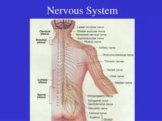
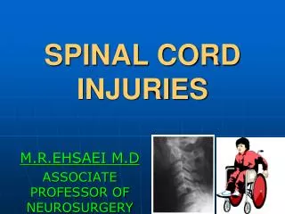
Anatomy of the Spinal Cord Structure of the spinal cord Tracts of the spinal cord Spinal cord syndromes . Spinal Cord. - Comparable to Input-Output (IO) System of the Computer - Spinal Nerve (C8, T12, L5, S5, Cx1) - Segmental Structure of Neural Tube Origin. Spinal segment

E N D
Anatomy of the Spinal Cord • Structure of the spinal cord • Tracts of the spinal cord • Spinal cord syndromes
Spinal Cord - Comparable to Input-Output (IO) System of the Computer - Spinal Nerve (C8, T12, L5, S5, Cx1) - Segmental Structure of Neural Tube Origin
Spinal segment C8, T12, L5, S5, Cx1 Anterior (Ventral) Root Posterior (Dorsal) Root Dorsal Root (Spinal) Ganglion Root - Rootlets
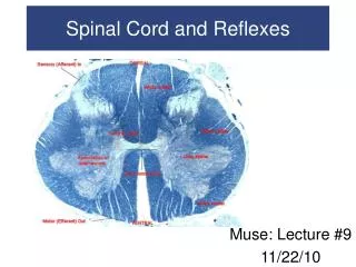
Spinal Cord External Figure Conus Medullaris (L1-2) Spinomedullary Junction - Foramen Magnum, Pyramidal decussation, C1 ventral root Enlargements - cervical (C5-T1) & lumbosacral (L1-L4) Longitudinal Fissures - anterior median fissure - anterolateral fissure - posterior median sulcus - posterolateral sulcus
Conus Medullaris (L1-2) Cauda Equina Anterior median fissure Anterolateral fissure
Posterior median sulcus Posterolateral sulcus Posterior intermediate sulcus Fasciculus cuneatus Fasciculus gracilis Posterior surface of the spinal cord
Spinal Cord Meninges Periosteum of Vertebra - Epidural Space ----------------- epidural anesthesia Dura Mater Spinalis Arachnoid Membrane - Subarachnoid Space -------- Lumbar Puncture Spinal Anesthesia Pia Mater Spinalis - Denticulate Ligament --------- Cordotomy - Filum Terminale
Meninges of • the spinal cord • Dura mater • Arachnoid membrane • Pia mater Denticulate ligament - specilization of the pia mater - landmark for cordotomy
Spinal Cord Vascular Supply Arterial Supply - Spinal Arteries Anterior (1) & Posterior (2) Spinal Artery from Vertebral artery - Radicular Arteries ----- Segmental arteries from Vertebral, Ascending Cervical, Intercostal and Lumbar Artery Venous Drainage - Longitudinal & Radicular Veins to Intervertebral veins ---- to Internal Vertebral Venous Plexus to external vertebral venous plexus ---- to segmental veins
5. Adamkiwicz artery anterior spinal artery segmental arteries
Spinal Cord Internal Structure White Matter Anterior Funiculus (Anterior White Column) Posterior Funiculus (Posterior White Column) Fasciculus Gracilis & Fasciculus Cuneatus Lateral Funiculus (Lateral White Column) Gray Matter Anterior Horn ------------ --- motor Posterior Horn -------------- sensory Lateral Horn ----------------- autonomic (sympathetic) Gray Commissure -------- anterior and posterior
Resembles a butterfly. • 2 lateral gray masses connected by the gray commissure. • Posterior projections are the posterior or dorsal horns. • Anterior projections are the anterior or ventral horns. • In the thoracic and lumbar cord, there also exist lateral horns.
Gray Matter • Posterior horns contain interneurons. • Anterior horns contain some interneurons as well as the cell bodies of motor neurons. • These cell bodies project their axons via the ventral roots of the spinal cord to the skeletal muscles. • The amount of ventral gray matter at a given level of the spinal cord is proportional to the amount of skeletal muscle innervated.
Gray Matter • Lateral horn neurons are sympathetic motor neurons serving visceral organs. • Their axons also exit via the ventral root. • Afferent sensory fibers carrying info from peripheral receptors form the dorsal roots of the spinal cord. The somata of these sensory fibers are found in an enlargement known as a dorsal root ganglion. • The dorsal and ventral roots fuse to form spinal nerves.
1. posterior horn 2. anterior horn 3. intermediate zone (intermediate gray) 4. lateral horn 5. posterior funiculus 6. anterior funiculus 7. lateral funiculus 8. Lissauer's tract 9. anterior median fissure 10. posterior median sulcus 11. anterolateral sulcus 12. posterolateral sulcus 13. Posterior intermediate sulcus
cervical enlargement (C8) thoracic cord (T8) lumbal enlargement (L3) sacral cord (S1)
Spinal Cord Internal Structure Principles of Cord Organization 1) Longitudinal Arrangement Fibers (White Matter) ------------- White Column Cell Groups (Gray Matter) ------- Gray Column 2) Transverse Arrangement Afferent & Efferent Fibers Crossing (Commissural and Decussating) Fibers 3) Somatotopical Arrangement
Spinal CordInternal Structure Laminae of Rexed Lamina I ---------- posteromarginal nucleus Lamina II ---------- substantiagelatinosa of Rolando Lamina III, IV ----- nucleus proprius Lamina V, VI Lamina VII --------- intermediate gray intermediolateral cell column (ILM) Clarke’s column (Nucleus dorsalis) intermediomedial cell column (IMM) Lamina VIII Lamina IX ---------- anterior horn (motor) cell Lamina X ----------- gray commissure
White Matter • Myelinated nerve fibers. • Allows for communication btwn the brain and spinal cord or btwn different regions of the spinal cord. • White matter on each side of the cord is divided into columns or funiculi. • Typically, they are ascending or descending. • What does that mean?
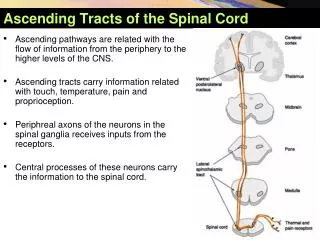
Spinal Cord Tracts Ascending Tracts Modality: Touch, Pain, Temperature, Kinesthesia Receptor: Exteroceptor, Interoceptor, Proprioceptor Primary Neuron: Dorsal Root Ganglion (Spinal Ganglion) Secondary Neuron: Spinal Cord or Brain Stem (Tertiary Neuron): Thalamus (Ventrobasal Nuclear Complex) Termination: Cerebral Cortex, Cerebellar Cortex, or Brain Stem
Spinal Cord Tracts Ascending Tracts Posterior White Column-Medial Lemniscal Pathway Spinothalamic Tract Spinoreticular or Spinoreticulothalamic Tract Spinocerebellar Tract Spinomedullothalamic Tract Cervicothalamic or Spinocervicothalamic Tract Spino-olivary Tract Spinotectal Tract
medial lemniscus lemniscal decussation internal arcuate fiber posterior white column posterior root Posterior White Column - Medial Lemniscal Pathway - ipsilateralloss of discriminative touch sensation and conscious proprioception belowthe level of lesion
spinothalamic tract anterior white commissure posterior root decussation Spinothalamic Tract - contralateral loss of pain and temperature sensation below the level of lesion
Descending tracts Lateral pathway • Voluntary movement of distal muscles • Direct cortical control Ventromedial pathway • Pose and antigravitational movements • Indirect cortical (stem) control
Corona Radiata lnternal Capsule, Posterior Limb Crus Cerebri, Middle Portion Longitudinal Pontine Fiber Pyramid Pyramidal Decussation Corticospinal Tract - Lateral and Anterior CR IC LPF Corticospinal Tract Pyr LCST PD - ipsilateralUMN syndrome at the level of lesion ACST
Spinal Cord Tracts ventromedial pathway dorolateral pathway Descending Tracts from Brain Stem
Spinal Nerves • 31 nerves connecting the spinal cord and various body regions. • 8 paired cervical nerves • 12 paired thoracic nerves • 5 paired lumbar nerves • 5 paired sacral nerves • 1 pair of coccygeal nerves
Spinal Nerves • Each connects to the spinal cord by 2 roots – dorsal and ventral. • Each root forms from a series of rootlets that attach along the whole length of the spinal cord segment. • Ventral roots are motor while dorsal roots are sensory.
The 2 roots join to form a spinal nerve prior to exiting the vertebral column. Roots are short and horizontal in the cervical and thoracic regions while they are longer and more horizontal in the sacral and lumbar regions. After emerging from its intervertebral foramen, a spinal nerve divides into dorsal ramus, ventral ramus, and meningeal branch that recurs to supply the meninges and associated blood vessels. Spinal Nerves
Each ramus is mixed. • Joined to the base of the ventral rami of spinal nerves in the thoracic region are the ramicommunicantes. These are sympathetic fibers that we’ll deal with shortly. • Dorsal rami supply the posterior body trunk whereas the thicker ventral rami supply the rest of the body trunk and the limbs.
Reflex Arcs • A reflex is a rapid, predictable motor response to a stimulus. Unlearned and involuntary. • Example? • Components of a reflex arc: • Receptor site of stimulus • Sensory neuron transmits afferent info to CNS • Integration center 1 or more interneurons • Motor neuron transmits efferent signals to effector • Effector muscle or gland
Reflexes • Reflexes involving skeletal muscles and somatic motor neurons are somatic. • Reflexes controlled by autonomic neurons are autonomic. • Spinal reflexes are integrated w/i the spinal cord while cranial reflexes are integrated in the brain. • Reflexes may be inborn or learned. • Reflexes may be monosynaptic or polysynaptic. • Difference?
Spinal Cord Syndrome • Predominantly Motor Syndromes • Poliomyelitis (Infantile Paralysis) • - viral infection of lower motor neuron • - LMN syndrome at the level of lesion • Amyotrophic Lateral Sclerosis (ALS) • - combined LMN and UMN lesion • - LMN syndrome at the level of lesion • - UMN syndrome below the level of lesion • - Lou Gehrig’s disease in USA
Spinal Cord Syndrome 1. corticospinal tract 2. lower motor neuron (LMN) Amyotrophic Lateral Sclerosis
Spinal Cord Syndrome Amyotrophic Lateral Sclerosis (ALS) Lou Gherig’s Disease Stephen Haking (1946- ) British Physicist, A Brif History of Time
Spinal Cord Syndrome Brown-Sequard syndrome (spinal cord hemisection) Major Symptoms 1. ipsilateralUMN syndromebelow the level of lesion 2. ipsilateralLMN syndrome at the level of lesion 3. ipsilateral loss of discriminative touch sensation and conscious proprioceptionbelow the level of lesion (posterior white column lesion) 4. contralateral loss of pain and temperature sensation below the level of lesion (spinothalamic tract lesion)
Spinal Cord Syndrome Brown-Sequard Syndrome (Spinal Cord Hemisection)