Download
1 / 29
310 likes | 683 Views
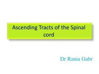
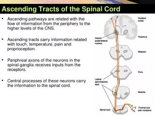
Spinal cord ascending tracts . Naming the tracts •If the tract name begins with “ spino ” (as in spinocerebellar ), the tract is a sensory tract delivering information from the spinal cord to the cerebellum (in this case)

E N D
Naming the tracts •If the tract name begins with “spino” (as in spinocerebellar), the tract is a sensory tract delivering information from the spinal cord to the cerebellum (in this case) •If the tract name ends with “spinal” (as in vestibulospinal), the tract is a motor tract that delivers information from the vestibular apparatus (in this case) to the spinal cord
•The three major sensory tracts involve chains of neurons •First-order neuron •Delivers sensations to the CNS •The cell body is in the dorsal or cranial root ganglion •Second-order neuron •An interneuron with the cell body in the spinal cord or brain •Third-order neuron •Transmits information from the thalamus to the cerebral cortex
Neurons in the sensory tracts are arranged according to three anatomical principles •Sensory modality •Somatotopic •Medial-lateral rule Sensory modality •Fine touch sensations are carried in one sensory tract •Somatotopic •Ascending tracts are arranged according to the site of origin •Medial-lateral rule •Sensory neurons that enter a low level of the spinal cord are more medial within the spinal cord •Sensory neurons that enter at a higher level of the spinal cord are more lateral within the spinal cord
The cell bodies of the 1st order neurons are located In the posterior root ganglion • lateral spinothalamic tract • pain, temperature • The pain impulses are transmitted to SC in fast-conducting delta A–type and slow conducting C-type fibers • The fibers of 1st order neurons terminate by synapsing with cells in the posterior gray column (substantiagelatinosa) • The axons of 2nd order neurons cross obliquely to the opposite side in the anterior gray and white commissures , ascending in the contralateral white column as the lateral spinothalamic tract • The lateral spinothalamic tract lies medial to the anterior spinocerebellar tract • Sacral fibers are lateral, cervical are medial • Pain fibers are slightly anterior to temperature • Ascends in medulla oblongata, pons, midbrain • ant spinothalamic tract + spinotectal + lateral spinothalamic = spinal leminiscus
The fibers of the tract end by synapsing with the 3rd- order neurons in VPL nucleus of thalamus • The axons of 3rd order neurons pass through internal capsule and corona radiata to reach the Post central gyrus of cerebral cortex
lateral spinothalamic tract Ventral nuclei in thalamus Midbrain Lateral spinothalamic tract 1st order neuron Second-order neuron Third-order neuron Pain and temperature sensations from right side of body
The cell bodies of the 1st order neurons are located In the posterior root ganglion • Anterior spinothalamic tract • crude touch and pressure • The fibers of 1st order neurons terminate by synapsing with cells in the posterior gray column (substantiagelatinosa) • The axons of 2nd order neurons cross obliquely to the opposite side in the anterior gray and white commissures , ascending in the contralateral white column as the anterior spinothalamic tract • Sacral fibers are lateral, cervical are medial • Ascends in medulla oblongata, pons, midbrain • ant spinothalamic tract + spinotectal + lateral spinothalamic = spinal leminiscus • The fibers of the tract end by synapsing with the 3rd- order neurons in VPL nucleus of thalamus • The axons of 3rd order neurons pass through internal capsule and corona radiata to reach the Postcentralgyrus of cerebral cortex
Anterior spinothalamic tract Anterior spinothalamictract Crude touch and pressure sensations from right side of body
Posterior white Column (Fasciculus gracilis, Fasciculus cuneatus ) Discriminative touch, vibratory sense, and conscious muscle-joint sense •Posterior Column tract consists of: •Fasciculus gracilis •Transmits information coming from areas inferior to T6 •Fasciculus cuneatus •Transmits information coming from areas superior to T6
The cell bodies of the 1st order neurons are located In the posterior root ganglion • The axons of 1st order neurons pass directly to the posterior white column • As Fasciculus gracilis and Fasciculus cuneatus • Fasciculus gracilis is present throughout the length of SC • Contains ascending fibers from sacral ,lumbar and lower six thoracic nerves • Fasciculus cuneatus is situated laterally in the upper thoracic and cervical segments • Is separated from Fasciculus gracilis by a septum • Contains ascending fibers from upper six thoracic nerves and all cervical nerves The fibers of Fasciculus gracilis and Fasciculus cuneatus ascend ipsilaterally and terminate by Synapsing on 2nd order neurons in nuclei gracilis and cuneatus of medulla oblongata
The axons of 2nd order neurons (internal arcuate fibers) cross the median plane (sensory decussation) Fibers then ascend as a single bundle (medial leminiscus) through medulla oblongata, pons, midbrain • The fibers of the tract end by synapsing with the 3rd- order neurons in VPL nucleus of thalamus • The axons of 3rd order neurons pass through internal capsule and corona radiata to reach the Postcentralgyrus of cerebral cortex
Posterior Columns Midbrain Medial lemniscus Nucleus gracilis and nucleus cuneatus Medulla oblongata Fasciculuscuneatus and fasciculusgracilis Fine-touch, vibration, pressure, and proprioception sensations from right side of body
The cell bodies of the 1st order neurons are located In the posterior root ganglion • Posterior spinocerebellar tract • muscle and joint sensation • 1st order neuron axons terminate at the base of post gray column (nucleus dorsalis) • the axons of 2nd order neurons enter posterolateral part of the lateral white matter • on the same side • ascend as the posterior spinocerebellar tract to medulla oblongata • Terminates in cerebellar cortex (through inferior cerebellar peduncle) • note: axons of lower lumbar and sacral spinal nerves ascend in the posterior white • column until they reach L3 or L4 segments where they synapse with nucleus dorsalis
The cell bodies of the 1st order neurons are located In the posterior root ganglion • Anterior spinocerebellar tract • muscle and joint sensation • 1st order neuron axons terminate at the base of post gray column (nucleus dorsalis) • the majority of axons of 2nd order neurons cross to opposite side and ascend as • anterior spinocerebellar tract in the contralateral white column • the minority of axons ascend as anterior spinocerebellar tract in the lateral white column • Of the same side • ascend as anterior spinocerebellar tract to medulla oblongata and pons • Terminates in cerebellar cortex (through superior cerebellar peduncle) • the fibers that crossed over in spinal cord cross back within cerebellum
Spinocerebellar Tracts PONS Cerebellum Medulla oblongata Anterior spinocerebellar tract Posterior spinocerebellar tract Spinal cord Proprioceptive input from Golgi tendon organs, muscle spindles, and joint capsules
The term dorsolateral fasciculus of the spinal cord refers to a bundle of nerve fibers located between the posterior white column and the lateral white column and bounded ventromedially by the posterior gray column of the spinal cord A longitudinal bundle of thin, unmyelinated and poorly myelinatedfibers capping the apex of the posterior horn of the spinal gray matter, composed of posterior root fibers and short association fibers that interconnect neighboring segments of the posterior horn
Regional variation of spinal cord structure cross sections taken from the cervical enlargement ( A ), midthoracic cord ( B ), and lumbosacral enlargement ( C )
cervical segment • an oval shape with large white matter funiculi • prominent, broad anterior gray horns that contain the motor neurons that innervate the upper extremities • thoracic segment • have a more rounded profile • exhibit small, slender, peglike anterior gray horns • lateral horns are unique to thoracic segments • lumbosacral segment • has very large anterior gray horns (motor supply to the lower extremities) • only a very small surrounding mantle of white matter
Cervical segment Fasciculus gracilis Fasciculus cuneatus Posterior white column Substantiagelatinosa Nucleus proprius Lateral white column Lateral group (UL) Accessory nucleus Medial group (neck) Anterior white column Anterior median fissure
Thoracic segment Fasciculus gracilis Fasciculus cuneatus Substantiagelatinosa Nucleus proprius Nucleus dorsalis Lateral horn (PREGANGLIONIC SYMPATHETIC) Motor neurons (trunk muscles)
Lumbosacral segment Fasciculus gracilis Substantiagelatinosa Nucleus proprius Lateral horn (PREGANGLIONIC PARASYMPATHETIC (S2-S3-S4) Motor neurons (LL muscles) Motor neurons (trunk muscles)