Download
1 / 33
390 likes | 564 Views
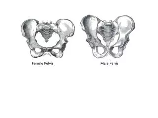
Female pelvis . Fetus as the object of labor . Obstetric terminology METHODS OF EXAMINATION. Measurement of pelvic spend by pelviometr . Usually measured four basic dimensions of the pelvis: three transverse and one d irect. The main dimensions of the pelvis. external conjugate.

E N D
Female pelvis. Fetus as the object of labor. Obstetric terminologyMETHODS OF EXAMINATION
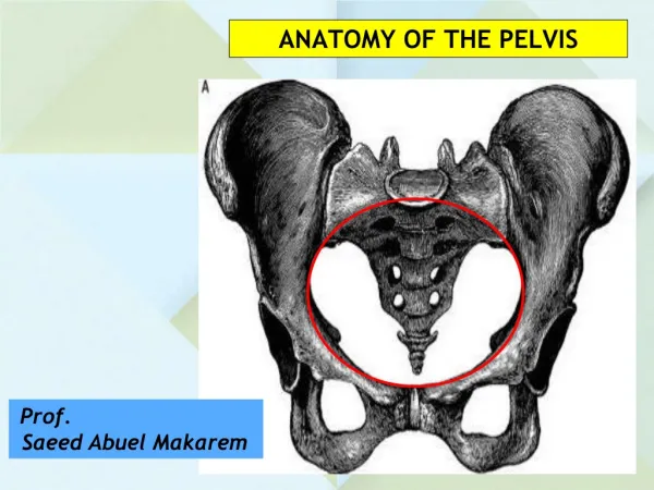
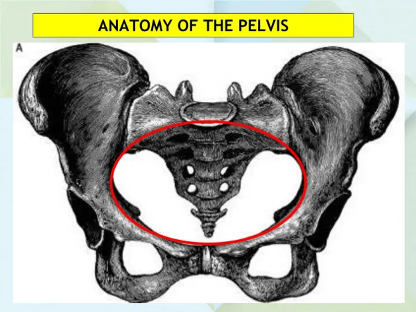
Measurement of pelvic spend by pelviometr.Usually measured four basic dimensions of the pelvis: three transverse and one direct.
external conjugate external conjugate – external size of pelvis. End of pelviometr set onmiddle of the upper margin of symphysis, the other end is over the sacral fossa contained between fifth lumbar vertebra and the beginning of the first sacral vertebra. External conjugate is20 cm
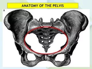
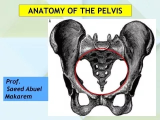
The planes of the pelvis and their dimensions • In obstetric practice are important dimensions of the pelvis, fromwhich depends on the course and outcomes for both mother and fetus. But most sizes pelvis can not be measured directly.Great pelvis for childbirth substantial does not matter, but in itssize may indirectly informates about the form and size of the pelvis.Pelvic cavity is the space between the walls, which the top and bottom limited inlet and outlet planes of the pelvis. It looks like a cylinder. In pelvic cavity are four planes:inlet, greatest dimention,narrow part (midpelvis) and outlet.
Small pelvis. The planes and the size of the pelvis • The line connecting the centers of all direct sizes of pelvis, called the main axis of the pelvis
Measuring the size of the pelvis • Rhombus of Michaelis - upper angle contained hollow under the spinous processes of the 5 lumbar vertebrae Lateral angles correspond posterior-superior iliac spine, lower- top sacrum. In women with a normal pelvis it has the correct form, approaching the square, its dimensions are 10.11 cm, height of the upper triangle 3-3.5 cm
Fetus as the object of labor • From all parts of the mature fetus most interesting head, because of the following reasons: 1) head has the big circumference and dense part of thefetus, which can withstand the greatest resistance from the birth canal and puts most pressure on them that determines the outcome of labor, 2) depending on the density and mobility of the cranial bones is greatly damage the birth canal of the mother and the fetus, and 3) the head of the fetus has a large number of cognitive items, which helps in diagnosing insertion and promotion in the bones of the pelvis.At the head of the fetus can distinguish two parts (Fig. 1): a relatively small front: lower jaw (1), maxilla (2) and very voluminous - brain. The latter consists of seven bones: two frontal (3), two parietal(4), one occipital (5), two temporal (6).
Fetus as the object of labor Sutures and fontanelles skull newborn (seen from above):1 - frontal suture, 2 - coronal suture 3 - sagittal (sagittal) suture 4 - occipital suture, 5 - Small fontanel 6 - large fontanel
Fetus as the object of labor 8. Direct size (diameter fronto-occipitalis) - from the nose to the occipital hill, length - 12 cm by 34 cm contours of equal 7. Average oblique size (diameter suboccipito-frontalis) - from suboccipital fossa to the anterior border of the scalp, length of 10 cm, and contours (circumferentia suboccipito-frontalis) - 33 cm 6. Small oblique size (diameter suboccipito-bregmatica) - from the middle suboccipital fossa large fontanel, length - 9.5 cm, and circumference suboccipito-bregmatica - 32 cm
Fetus as the object of labor • Attitude of fetus (habitus) - is the ratio of the limbs of the fetus and the head to his body. In the most favorable habitus - curved spine, resulting in back arched outwards, head bent, chin close to the chest. The legs are flexed at the hip and knee joints, intersect and pinned to the lower abdomen. Handles are flexed at the elbows and intersect on his chest.
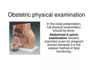
METHODS OF EXAMINATION • Ultrasonic dating of the pregnancy and an ultrasonic fetal survey to detect gross abnormalities have been recommended in some clinics as a routine part of early prenatal care. Routine ultrasonography is most cost – effective in patients in whom the date of the last menstrual period is uncertain and in patients with a family history of congenital anomalies. Considerable individualization should be exercised in making the decision to order this evaluation. If ultrasonography is performed, it is most informative between 11-13 and18-20 weeks.
METHODS OF EXAMINATION Auscultation. In cephalic presentation, the point of maximal intensity of fetal heart sounds is usually midway between the maternal umbilicus and the anterior-superior spine of her ilium