Download

1 / 28
300 likes | 616 Views
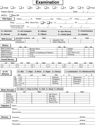
METHODS OF EXAMINATION. • Biographical details • Medical history • Chief complaint • History of present complaint • Dental history • Social history • Extraoral examination • Intraoral examination • Special tests. 1. Have you ever had Rheumatic Fever? Yes No

E N D
• Biographical details • • Medical history • • Chief complaint • • History of present complaint • • Dental history • • Social history • • Extraoral examination • • Intraoral examination • • Special tests
1. Have you ever had Rheumatic Fever? • Yes • No • 2. Do you have Heart Trouble or High Blood Pressure? • Yes • No • 3. Do you have Chest Trouble? • Yes • No • 4. Have you had Jaundice or Hepatitis, or been refused as a blood donor? • Yes • No • 5. Have you ever had severe bleeding that needed special treatment after an • injury or dental extraction? • Yes • No • 6. Is there any family history of Bleeding Disorders? • Yes • No • 7. Are you taking any Drugs, Tablets, or Medicines? • Yes • No
8. Do you suffer from any Allergies (e.g. Penicillin)? • Yes • No • I f 'Yes' please list • 9. Are you Diabetic? • Yes • No • 10. Do you have any history of Epilepsy? • Yes • No • 11. Have you had any a) Serious Illnesses or Operations? • Yes • No • or b) Adverse reactions to Local or General Anaesthesia? • Yes • No • 12. Have you come into contact with anybody who has AIDS or is HIV positive? • Yes • No • 13. (Females only) Are you pregnant?
CHIEF COMPLAINT This is the opportunity for the general practitioner to let the patient describe a dental problem as it appears to him/her. You may start with 'Tell me about your problem' or 'How can I help?' Allowing time to listen to the patient in a busy schedule can pay dividends in reaching the correct diagnosis swiftly and avoiding embarrassing mistakes. A distressed patient will be put at ease, and conversation can then lead into more detailed discussion
HISTORY OF PRESENT COMPLAINT • When did the pain or problem start? • Does anything make the pain better orworse? • Relieving factors. • The frequency of painful episodes. • Intensity. • Location. • Duration. • Postural changes. • Does anything trigger the pain? • Quality of pain.
Facial Swelling Are there any signs of acute inflammation - heat, swelling, redness, pain, loss of function and does thepatient have a raised bodytemperature?Does the patient feel that his/her face isswollen in any way? Ask patients to look in amirror and point to any perceived swelling. The practitioner can assess the facial contour in profile and by looking down the bridge of the nose from above to see any asymmetry inthe nasolabial folds. Facialasymmetry can be due to guarding of painfultissues.
Asymmetry in the right nasolabial folds is more visiblewhen viewed from above.
Palpation Lymph nodes can be gently palpated with thefingertips. Lymphadenopathy of the submandibularlymph nodes could be an indicationof infection in the oral cavity. Tendernessmay indicate a site of acute inflammationdeep to the skin
Palpation of the submandibular lymph nodes. The clinicianis positioned behind the patient and palpates thenodes gently with finger tips.
Is it possible for the patient toopen his/her mouth sufficiently wide for rootcanal treatment? If two fingers can be placedbetween the maxillary and mandibular incisortips then it should be possible to instrumentmost teeth
Sufficient opening is required to gain access to the teethfor endodontic treatment. Two fingers' width in thei ncisor region is perfectly adequate.
General condition of the mouth: • Is the mouthin good health or neglected? Are there heavyplaque deposits and evidence of gross periodontaldisease? Are restorations ofgood quality, or are the margins overhangingand poorly finished? Is there obvious recurrentcaries present
A neglected mouth. The patient will need advice on oralhygiene prior to endodontic treatment.
Tooth mobility: • A suspect tooth can bemoved gently by finger and thumb pressure;any horizontal mobility is then gradedMobility can result from trauma, root fractures, periodontal disease and gross rootresorption. Sometimes a very slight (< 1 mm)degree of mobility may be normal. Forinstance, a tooth that has a horizontal rootfracture in the middle third could be expectedto have a degree of mobility, as would teeth under active orthodontic traction. Neitherwould necessarily require treatment purelybecause of the mobility.
Testing tooth mobility by gently applying lateral forcesbetween finger and thumb.
Tenderness to palpation: • The tooth is movedvertically and side to side with finger pressure.Teeth with acute apical periodontitiswill often be tender when palpated in thismanner.
Percussion: • Tapping a tooth with a mirrorhandle can help identify replacement resorption(ankylosis). A characteristic ringingsound is sometimes heard on percussion
Gently percussing a tooth with a mirror handle may elicitthe classical ringing sound that occurs with replacementresorption (ankylosis).
Palpation of the buccal sulcus: • Running afinger gently along the buccal sulcus will helpelicit if there is any swelling or tendernessover the apex of an offending tooth
Palpating the buccal sulcus over the apices of the teeth,with a finger tip. Any tenderness or swelling is noted.Tenderness may be an indication of acute apical periodontitis.
Periodontal pocketing: • Probing depths shouldbe measured carefully with a periodontalprobe. Ideally a probe with a tip of 0.5 mmshould be used and pressure of no more than25 g applied (light pressure!). Broad pocketsare normally due to periodontal disease. Asudden increase in probing depth resulting ina narrow but deep pocket may indicate theposition of a vertical root fracture or sinustract lying within the periodontal ligament
The maximum periodontal probing depth on the mesialaspect was 7mm. The pocket shape was deep and narrow.
The probing depth of 7mm on the distal aspect of thetooth directly opposite to that on the mesial aspect wasindicative of a vertical root fracture.