Download

1 / 31
310 likes | 535 Views
SKIN AND APPENDAGES OF SKIN. Dr Iram Tassaduq. THE INTEGUMENT. The integument is the body’s most massive organ. It is composed of skin that covers

E N D
SKIN AND APPENDAGES OF SKIN DrIramTassaduq
THE INTEGUMENT • The integument is the body’s most massive organ. • It is composed of skin that covers the entire body, together with certain accessory organs which are derivatives of the skin, such as nails, hair, sweat and sebaceous glands.
ESSENTIAL FUNCTIONS • Acts as a barrier • Provides immunologic information • Participates in homeostasis • Conveys sensory information • Performs endocrine functions • Functions in excretion
1. STRATUM BASALE/ STRATUM GERMINATIVUM • Single layer of cells • Cuboidal / Columnar cells • Closely spaced nuclei • Basophilic cytoplasm • Contains melanin • Cell junctions • Contains stem cells • Provides epidermal cell renewal
2. STRATUM SPINOSUM • Several cells thick • Larger cells • Cytoplasmic processes or spines • Desmosomes • Prickle cells • Flattening of cells towards surface
3. STRATUM GRANULOSUM • Most superficial layer of nonkeratinized portion of epidermis • One to three cell layers thick • Keratohyalin granules
4. STRATUM LUCIDUM • A subdivision of stratum corneum • Normally well seen in thick skin • Refractile appearance • Stains poorly • Eosinophilic cells
5. STRATUM CORNEUM • Cells are flattened, desiccated, anucleated • Most differentiated cells in the skin • Filled almost entirely with keratin filaments • Thick plasma membrane • Water barrier in epidermis • Variation in the thickness of layer
Dermis varies from 0.2 to 4 mm in thickness • Composed of dense, irregularly arranged C.T. • Contains three types of C.T. fibres plus fibroblasts and macrophages • Two layers can be distinguished • Junction between dermis and epidermis
LAYERS OF DERMIS 1. THE PAPILLARY LAYER • Consists of loose C.T. • Thickness of collagen fibres • Contains type I and type III collagen • Irregular network of elastic fibres • Dermal papillae and ridges • Blood vessels • Nerve processes
THE RETICULAR LAYER • Always considerably thicker and less cellular than the papillary layer (although its thickness varies) • Characterized by thick irregular bundles of mostly type I collagen and by coarse elastic fibres • Langer’s lines
THIN HAIRY SKIN THICK NONHAIRY SKIN
FLEXOR OR JOINT LINES • Major markings found in the vicinity of synovial joints where the skin is attached strongly to underlying deep fascia. • Prominent on the flexor surfaces of palms, soles and digits. • Skin lines don't necessarily coincide with the underlying joint line.
PAPILLARY/EPIDERMAL/FRICTON/RIDGES • A friction ridge is a raised portion of the epidermis on the fingers and toes, the palm of the hand or the sole of the foot.
WRINKILE LINES • Caused by contraction of underlying muscles, present perpendicular to their axis of shortening. • On face, they are known as lines of facial expression, aging makes them permanent due to loss of skin elasticity.
BLOOD SUPPLY OF SKIN • The dermis contains horizontally arranged superficial and deep plexuses, which are interconnected via communicating vessels oriented perpendicular to the skin surface.
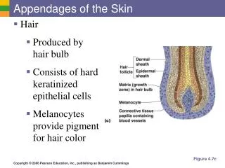
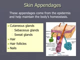
APPENDAGES OF SKIN • Nails • Hair follicles • Sweat glands • Sebaceous glands
HAIR • Hairs are elastic keratinized threads that develop from epidermis • Has free shaft and root embedded in skin • Length: 1 mm. to 5 feet • Humans: hair covers entire body except: palm, sole, region of anal & urogenital apertures
NAILS • The nails are horny plates that form a protective covering on the dorsal surface of the terminal phalanges of the fingers and toes
SWEAT GLANDS(SUDIFEROUS GLANDS • widely distributed on body • Sweat – is a blood filtrate • 99% water with some salts • Contains traces of metabolic wastes • Vital for thermoregulation • Also influence water and ion balance
SEBACOUS GLANDS • One to several sebaceous glands always connect with a hair • A gland usually is located in the angle between the follicle and its muscle • Its short duct empties into the follicle at a level three-fourths of the way up.
SEBACOUS GLANDS • Occur over entire body • Except palms and soles • Secrete sebum – an oily substance • Simple alveolar glands • Most are associated with a hair follicle • Functions of sebum • Collects dirt; softens and lubricates