Download
1 / 152
1.53k likes | 1.58k Views
Explore the detailed structure of the skin, including the epidermis, dermis, and subcutaneous layer. Learn about different cell types, layers of the epidermis, and characteristics of thin and thick skin. Understand the role of epidermal growth and repair for maintaining healthy skin.

E N D
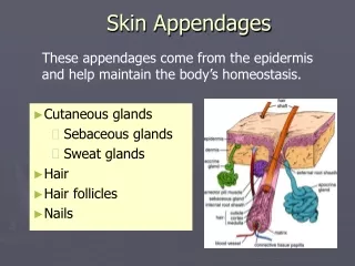
Structure • Picture page 163 ( 197) • Integumentary system = skin • Skin is a thin relatively flat organ • Classified as a membrane: cutaneous membrane • Cutaneous Membrane is divided into epidermis and dermis
Epidermis • Outer, thinner epithelial layer. • Develops from ectodermal germ layer, usually by the 17th week of gestation the baby’s has all the essential characteristics of the adult’s • Avascular
Dermis • Inner, thicker connective layer • Derived from the mesoderm • Vascular
Dermal-epidermal junction • Specialized area where cells of epidermis meet connective tissue of dermis • characteristics of the adult structure by the 9th week of gestation
Subcutaneous Layer • Also called hypodermis • Lies beneath the dermis • Rich in fat and aerolar tissue • Irregular connective tissue
Epidermis Dermis Subcutaneous layer (hypodermis)
Epidermis Page 200 in home books
Cell Types • Epidermis is composed of several types of epithelial cells • Keratinocytes • Most important cell in the epidermis • Comprise over 90% of epidermal cells • Filled with tough, fibrous protein called keratin • principle structural element of outer skin
Cell Types • Melanocytes • Contribute color to skin • Protect from UV light • Can be completely absent from skin in some non-lethal conditions
Cell Types • Langerhans Cells • Dendritic cells (immune cells) • Play a role in immune reactions that effect the skin • Cells originate in the bone marrow but migrate to deep cell layers of the epidermis early in life
Cell Layers of EpidermisStrata Page 200 in home books
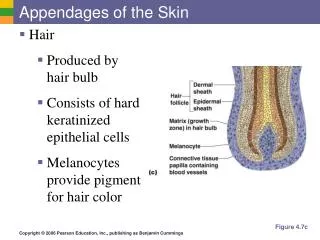
Stratum basale • Base layer • Single layer of columnar cells • Only cells in this deepest layer of epithelium undergoes mitosis • Cells migrate from this layer to other layers until they reach the surface and are shed off
Stratum Spinosum • Also called spiny layer • Stratum germinativum is used to describe the stratum basale and Stratum Spinosum together • Formed from 8-10 layers of irregularly shaped cells with very prominent intercellular bridge or desmosomes • Desmosomes appear to pull points of the plasma membranes of adjoining cells toward one another. Gives spiny appearance • Cells are rich in RNA making them well equipped to start protein synthesis needed for the production of keratin
Stratum Granulosum • Granular layer • Process of surface keratin formation begins • Sheet 2-4 layer deep filled with intensely staining granules called keratohylin (required for surface keratin formation) • Cells start to degenerate • High levels of lysosomal enzymes are present in cytoplasm and nuclei are in the process of breaking down • In thin skin this layer may not be visible
Stratum Lucidum • Clear layer • Keratinocytes are very flat, closely packed and clear • Nuclei are usually absent • Dying cells are filled with eleidin which is eventually transformed to keratin • Absent in thin skin
Stratum Corneum • Horny layer • Most superficial layer of epidermis • Composed of thin squamous cells • At surface cells are dead and continuously being shed • Desmosomes holding together Keratinocytes strengthen this layer • Keratinization: process in which cells from deeper layers migrate, fill with keratin and move to surface
Stratum Corneum • Sometimes called barrier area of skin • Protects from water loss and environmental threats • Glycophospholipids cement keratin into water proof barrier. • Glycophospholipids can be washed away by excessive soaking. The keratin can then absorb water appearing puffy and wrinkled • Diseases can cause layer to thicken • Hyperkeratosis: Thick, dry, scaly skin that is inelastic and subject to painful fissures
Thin and Thick Skin • There are up to 5 layers of stratum or cell layers. (stratum corneum, lucidum, granulosum, spinosum, basale) • Epidermal tissue can be categorized thin or thick skin • Most of the body surface is covered by thin skin • Hairless skin covering palms, fingertips, soles of feet or other areas associated with friction has a covering of thick skin
Thick skin • Each of 5 strata of epidermis are present and each stratum are generally several layers thick • Hair not found in thick skin • Thick skin underlying dermal papillae are raised in curving parallel friction ridges forming fingerprints or footprints • Ridges allow us to pick up and manipulate small items and supplies slip resistance to the feet.
Thin skin • Number of cell layers in the epidermal stratum are less than in thick • One or more strata may be entirely absent • Friction ridges are not present
Epidermal Growth and Repair • Turnover or regeneration time describes period required for a cell population to mature and reproduce • To maintain constant thickness, new cells must be formed at the same rate that old keratinized cells flake off from stratum corneum. • Current research suggest regeneration time is about 35 days • Abrasion can accelerate skin regeneration time. The result is an intense stimulation of mitotic activity in the stratum basale and shortened turnover period • Continued abrasion can result in an abnormally thick stratum corneum, producing calluses at the point of abrasion.
Epidermal Growth and Repair • Callus formation is normal but there are several skin diseases by abnormally high mitotic activity in the epidermis resulting in scales and lesions • 10%-12% of cells in the stratum basale enter mitosis each day • Cells migrating to surface proceed upward in vertical columns from groups of 8-10 of these basal cells undergoing mitosis • Each group of active basal cells, with its vertical column of migrating keratinocytes is called epidermal proliferating unit (EPU)
Review • Identify two main layers of skin • Terms thick and thin refer to which primary layer of skin • How do thin and thick skin differ • Identify two main cell types found in the epidermis • List 5 layers of epidermis (strata)
Dermis Page 200 in home books
Dermal-Epidermal Junction • Specialized area where cells of epidermis meet connective tissue of dermis • characteristics of the adult structure by the 9th week of gestation • lies between papillary layer and the stratum basale • Combines basement membrane and includes specialized fibrous elements and a polysaccharide gel • cement the superficial epidermis to the dermis (glue the two layers together) • Provides mechanical support for epidermis
Dermis • Also called the corium or “true skin” • Vascular • Composed of thin papillary layer and thicker reticular layer • Much thicker than the epidermis • Serves a protection function against mechanical injury
Dermis • Specialized network of nerves and nerve endings called somatic sensory receptors process sensory information • At various levels the dermis may contain muscle fibers, hair follicles, sweat and sebaceous glands and many blood vessels • It is rich vascularity that plays an important role in temperature regulation (discussed later)
Papillary Layer • Thin superficial layer of the dermis • Forms bumps called dermal papillae on its surface • Dermal-Epidermal Junction lies between papillary layer and the stratum basale • Composed of loose connective tissue elements along with thin collagenous and elastic fibers • Has characteristic ridges on surface due to conforming tightly to dermal papillae • Fingerprints and footprints allow us to grip so we can grasp small objects and walk upright on slippery surfaces (Friction Ridges)
Reticular Layer • Thicker layer of dermis • More of the dense reticulum or network of fibers (most are collagenous) than in papillary layer • Dense collagenous fibers in this layer is what produces leather in processed animal skins (these fibers also give skin toughness) • Elastic fibers make skin stretchable
Reticular Layer • Serves as a point of attachment for skeletal and smooth muscle fibers • Most of the structures such as muscle fibers, hair follicles, sweat and sebaceous glands are located in reticular layer • Several skeletal muscle are located in the skin of the face and scalp and permit various facial expressions
Reticular Layer • Distribution of smooth muscle is much more extensive than skeletal • Each hair follicle has a small bundle of involuntary muscle attached to it. Called arrector pili muscle • Contraction of these muscles makes hair stand on end and raises skin around hair • Goosebump • Have millions of somatic sensory receptors
Dermal Growth and Repair • Unlike epidermis, dermis does not continually shed and regenerate • Rapid regeneration of connective tissue in dermis occur only unusual circumstances such as wound healing • Fibroblasts quickly reproduce and begin forming a dense mass of new connective tissue that will either be replaced by normal tissue or become a scar
Dermal Growth and Repair • Dense white bundles of collagenous fibers orient themselves in patterns called cleavage lines or Langer’s Lines. Page 167 (203) • Surgical incisions are made parallel to cleavage lines the resulting wound has less tendency of gaping open and will tend to heal with a less noticeable scar • When elastic fibers are stretched too much (pregnancy) Fibers weaken and tear and initially resulting in pinkish or slightly bluish depressed furrows with jagged edges • When they heal and lose color they remaining furrows appear as glistening silver-white scar lines (stretch marks)
Hypodermis • Called subcutaneous layer, subQ or superficial fascia • Not part of the skin but usually discussed with the skin • Forms connection between skin and underlying structures of the body • Made up mostly of loose fibrous tissue and adipose tissue • Carries major vessels and nerves to skin above
Review • What is the name of the layer separating the dermis from the epidermis? • Which layer of the dermis forms the bumps that produce the ridges on palms and soles? • Which layer is vascular? Dermis or epidermis • What is the main function of the hypodermis?
Melanin • Main determinant of skin color is the quantity of melanin deposited in the epidermis cells • Melanin (pigment) is produced by melanocytes • Melanocytes scattered throughout the stratum basale is roughly the same amount for everyone • It is the amount and type of melanin pigment that melanocytes produce that account for skin color variations
Melanin • Two groups of melanin • Eumelanin • Pheomelanin • Eumelanin • “true Black” • Very dark brown, sometimes nearly black • Dark skinned and dark haired produce large quantities • Absorbs more UV radiation than pheomelanin