Download
1 / 52
520 likes | 878 Views
Benign Disease of the Breast. Dr. Bader Ashkanani Dr. Ibtisam Al-Bader. Objectives. Anatomy of the female breast Development and Physiology of the female breast Diagnostic approach Common Benign disease of the breast. Anatomy. Modified sweat glands

E N D
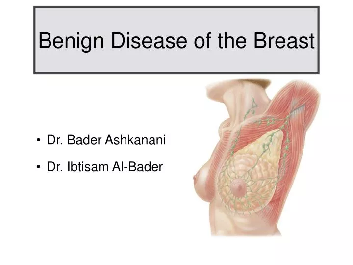
Benign Disease of the Breast • Dr. Bader Ashkanani • Dr. Ibtisam Al-Bader
Objectives • Anatomy of the female breast • Development and Physiology of the female breast • Diagnostic approach • Common Benign disease of the breast
Anatomy • Modified sweat glands • Lie in superficial fascia of ant. chest wall • Ant. To pectorals major with retromammary in between • Clinically devided into 4 quadrants • Nipple-areola complex • Axillary tail = Tail of Hence
Anatomy • Composed of 2 types of tissue: • - glandular : lobules ( produces milk) & ducts (carry milk to nipple) • - stromal: connective tissue supports the breast ( ligaments of Cooper)
Blood supply • Perforating branches: • - internal thoracic artery • - intercostal arteries • - lateral thoracic artery • - thoracoacromial artery • Veins correspond to art.
Lymph Nodes • Clinical: medial, lateral, anterior, posterior, apical • Anatomical: lateral, anterior (pectoral), posterior (subscapular), central, subclavicular, interpectoral (Rotter's) • Relation to pectoralis minor Msl. • - Level I : below and lateral • - Level II : deep to Msl. • - Level III : above and medial
Applied Anatomy • Long thoracic nerve: • - supply serratus ant. • - injury -> winged scapula • Thoracodorsal nerve: • - post. cord of brachial pl. • - supply latissmus dorsi • - injury -> medial rotation, adduction and extension of shoulder joint • Medial and lat. Pectoral nerves • - supply pact. major • - injury --> adduction and flexion of shoulder
Objectives • Anatomy of the female breast • Development and Physiology of the female breast • Diagnostic approach • Common Benign disease of the breast
Fetal and Neonatal • Linear thickening of ectoderm at 6th week known as the milk ridge • Ridge extends from axilla to inguinal region • Disappear giving rise to breast parenchyma • Localized depression in the line --> nipple • Areola recognized at 5 months
Fetal and Neonatal • Witch's milk: • Milk expressed from the breasts of both sexes in majority of newborns due to abrupt withdrawal of maternal hormones • May also be associated wit breast hypertrophy ( physiological gynecomastia )
Childhood • Minimal branching of the ducts • No alveolar formation • End of it --> Puberty
Puberty • GnRH --> FSH & LH --> estrogen & progesterone • Estrogen: ductal growth and branching • Progesterone : development of lobular units • Increase in periductal connective tissue
Pregnancy • Intense stimulation of breast • Hormonal drive : sex steroids, HCG, HPL, prolactin and growth hormone • Two major phases: developmental & differentiation
Phase I - Developmantal • Increase in numbers of alveoli • Marked ductal elongation • Engorged, heavy breasts • Darkening of nipple and areola
Phase II - Differentiation • Under progesterone influence • Differentiation of lobules into secretory units • Massive increase in blood supply • Breast size and weight increase
Menopause • Cessation of ovarian function • Age of 50's • Reduction in numbers of ducts and lobules • Breast tissue replaced by fat deposition
Objectives • Anatomy of the female breast • Development and Physiology of the female breast • Diagnostic approach • Common Benign disease of the breast
Diagnostic approach • History & Physical Examination • Biopsy • Imaging
History • History of the mass • Constitutional symptoms if Ca suspected • Age at menarche/menopause • Pregnancies & lactation • HCT & OCP • Oopherectomy • Past & family history
Physical Examination • Upright sitting • Inspection • Palpation • Lymph nodes
Imaging • Detect small, non palpable breast abnormality • Evaluate clinical findings & guide diagnostic procedures • Mammogram - MRI - Ultra sound (U/S)
Mammogram • Best for screening • Best in women above 35 yrs • Standard vs. Digital mammo. • BI-RADS scoring
Breast Imaging Reporting and Data System (BI-RADS) • - CATEGORY DEFINITION: • 0 Incomplete assessment—need additional imaging evaluation or prior mammograms for comparison • 1 Negative—nothing to comment on; usually recommend annual screening • 2 Benign finding—usually recommend annual screening • 3 Probably benign finding (<2% malignant)—initial short-interval follow-up suggested • 4 Suspicious abnormality (2%-95% malignant)—biopsy should be considered • 5 Highly suggestive of malignancy (>95% malignant)—appropriate action should be taken • 6 Known biopsy—proven malignancy
MRI • Evaluate primary tumor + extension • For young women • Pre-op for breast conservation • Not useful in screening • Sensitive for invasive Ca, not DCIS
Ultrasound • Cystic vs solid • Tense breast • Operator dependent • False positives ( Berg WA, 2008 ) • Not effective in screening
Biopsies • Fine Needle Aspiration Biopsy ( FNA ) • Core Needle Biopsy
FNA • 22 gauge needle, syringe, 95% alcohol prep./ Normosol • Differ. cystic/solid, but used in new masses • Diagnostic ( cytology ) & therapeutic ( aspiration ) • May require further core biopsy according to FNA result
Core Needle Biopsy • Done under U/S, mammo., MRI guidance • Patient in prone position • Multiple samples through small incision usin 11 gauge needle with vacuum assistance • Clip is placed to localize the lesion • Provide grading and receptor status
The diagnosis of breast lesions using a minimally invasive procedure, such as core needle biopsy, is the preferred approach. The use of excisional breast biopsy as a diagnostic procedure increases costs and results in delays to definitive surgery for patients with cancer. • Nelson H, Tyne K, Naik A, et al: Screening for breast cancer: Systematic evidence review update for the U.S. Preventive Services Task Force. Ann Intern Med 2009
Non-palpable abnormalities • Detected by mammography • Further investigation according to BI-RADS • Image guided core biopsy in 75 - 80 % of cases • Wire-Localized surgical excision
Wire-Localized excision • Diagnosis not yet obtained / ADH --> DCIS • Wire through introduced needl • Under mammography guidance • Nearby lesion
Objectives • Anatomy of the female breast • Development and Physiology of the female breast • Diagnostic approach • Common Benign disease of the breast
Fibroadenoma • Commonest tumor < 30 yrs • Firm, mobile, smooth / lobulated, may increase in size • Related to high estrogen level • Benign Tumor, rarely neoplastic • Simple vs complex
Fibroadenoma • Dx : • - Ultrasound • - FNAC --> stromal & epithelial elements • Tx : • - Reassurance • - Surgical excision • - Cryoabillation
Papillomas • Solitary intraductal papilloma • Often close to areola, but can occur peripherally • < 1 cm, can increase up to 4 cm • Causes bloody nipple discharge • No high risk of Ca • Tx : surgical excision
Breast cysts • Very common ( age 30 - 40 ) • Smooth, firm, mobile, +\- tenderness, anywhere in breast • Rarely malignant - disappear at menopause • Associated with OCP
Radial scars • Complex sclerosing lesions • Contain microcysts, epithelial hyperplasia, adenosis and central sclerosis • Architectural distortion on mammogram • Associated with increased risk of Ca • Tx is surgical excision to r/o Ca
Sclerosing adenosis • Proliferation of stromal tissue • Microcalcification on mammogram • Component of fibrocystic disease • No malignant potential
Phyllodes Tumor • Large, fast growing masses • < 1% of breast neoplasia • Arise from periductal stroma • Can be benign, borderline, malignant • Occur in adult women (40-50) • Composed of both epithelial & stromal element
Phyllodes Tumor • Tx: surgical excision + safety margin • Chemotherapy , radiotherapy , hormonal therapy doesn't work much • Regular imaging and f/u necessary
Breast infections and abscess • Lactational infections • Chronic subareolar infections ( duct ectasia )
Lactational infections • Fever, leukocytosis, erythema, tenderness • Most common --> Staph. Aureus • Mastitis --> Tx: antibiotics + frequent emptying of the breast • Abscess --> Tx: surgical drainage + antibiotics
Chronic subareolar infections • Non lactating --> subareolar ducts infected • Periductal mastitis / Duct ectasia • Mixed: aerobic + anaerobic skin flora • Associated with smoking and diabetes
Chronic subareolar infections • Series of infections • - Inflammatory changes and scarring • - Retraction / inversion of nipple • - Chronic fistulas from subareolar ducts to periareolar skin • - Subareolar masses ( on palpation & mammo. )
Chronic subareolar infections • Tx: • - Early --> antibiotic + warm soaks • - Abscess --> drainage + antibiotics • - Recurrent --> excision of entire subareolar duct complex after infection resolves + I.V. Antibiotic • - Rarely, excision of nipple and areola
Gynecomastia • Hypertrophy of breast • Most common benign disease of male breast • Increase in both stromal and ductal elements • Unilateral / Bilateral • Physiological vs. Pathological
Gynecomastia • Pathological • - Drugs: digoxin , diuretics , marijuana , heroin , TCA , anabolic steroids • - Disease: liver disease , bronchioectasis , T.B. , COPD, testicular tumors • Tissue biopsy to exclude malignancy • Tx: surgical excision
Galactocele • Milk-filled cyst • Occur after cessation of breast feeding • Center of breast or below nipple • Pathogenesis unknown • Aspiration --> thick creamy fluid • Surgery in infected / difficult to aspirate cysts