Benign Breast Mass
Benign Breast Mass. Three females with ages 23, 35, and 55 years, respectively, went to see you for consult. All have a mass in one of their breasts. . A. What important general data from the patients do you think are important to be able to guide you in your diagnosis?. Age


Benign Breast Mass
E N D
Presentation Transcript
Three females with ages 23, 35, and 55 years, respectively, went to see you for consult. All have a mass in one of their breasts.
A. What important general data from the patients do you think are important to be able to guide you in your diagnosis? • Age • Risk increases with age • Gynecologic History: • Menarche • Menopause (age 55) • Parity • Early full term pregnancy is protective • Multigravida is protective • Breastfeeding practices • Breast Feeding protective • Use of HRT, oral contraceptives
A. What important general data from the patients do you think are important to be able to guide you in your diagnosis? • Family History (Cancer) Institute of Public Health UKRelative Risk of Ca • Other Risk Factors • History of radiation exposure • Obesity
B. In the Physical Examination, differentiate a benign from a malignant lesion. It is not possible to distinguish a benign from a malignant cyst by physical exam alone with certainty. Benign Lesion Malignant Lesion Firm Indistinct borders Attchments to the skin or deep fascia with dimpling or nipple retraction Bloody discharge from nipple Skin changes on breast (redness, crusting, dimpling) • Freely movable • Regular edges • Rounded feeling • More likely to feel tender to touch
C. How will you approach a 23-year old, with a 2 X 2 X 2cm, firm, mobile, well-circumscribed non-tender mass on her L breast? • Diagnosis? • Role of imaging modalities for this case. • Options in the management?
Fibroadenoma • Benign • Common in young females (20-30 years old) • Usually small (2-3cm) • Related to estrogen • Not premalignant
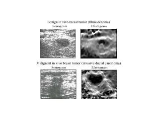
Ultrasound • A small circumscribed discrete mass suggests the presence of a simple cyst or fibroadenomafor women younger than 30 years old • Distinguish between cystic and solid masses • Not an effective screening test for cancer • Can confirm the diagnosis of a simple cyst or support a diagnosis of fibroadenoma • Can diagnose a simple cyst if four criteria are fulfilled: • Round or oval shape • Sharply defined margins • Lack of internal echoes • Posterior accoustic enhancement
Ultrasound of Fibroadenoma • Well-circumscribed elliptic mass • Uniform echogenicity • Lesion is larger in the transverse than in the anteroposterior direction • Well-demarcated margins
D. 35-year old, with a 2 x 2 x 2cm, firm, mobile, well-circumscribed non-tender mass on her R breast • Role of imaging modality? Choice? • A mammogram was taken as seen in the picture. Is this benign or malignant? • Differentiate radiologically a benign lesion from a malignant one. • Should the patient have a mother who is a breast cancer survivor, how would that information change your management?
Role of Imaging Modalities • Diagnostic Mammography • To screen the normal surrounding breast tissue and the opposite breast for non-palpable cancers • To make a diagnosis of the palpable mass
A mammogram was taken as seen in the picture. Is this benign or malignant? Normal Benign
Differentiate radiologically a benign lesion from a malignant one. Benign lesion • round or oval smooth-edged masses with the outline clearly defined
Differentiate radiologically a benign lesion from a malignant one. Malignant • Spiculated density with ill defined margins • Suggestive features include: • Clustered microcalcifications • Asymmetric density • Ductalassymetry • Distortion of skin, nipple and normal breast architecture There is a small spiculatedtumour in the middle of the right breast. Note the asymetry.
Should the patient have a mother who is a breast cancer survivor, how would that information change your management? • Close surveillance with SBE, mammography and possibly MRI • SBE at age 18 • Semi-annual SBE at age 25 • Annual mammography beginning age 25 or 10 yrs prior earliest age of onset of a family member • Chemoprevention using Tamoxifen