Download
1 / 26
270 likes | 2.03k Views
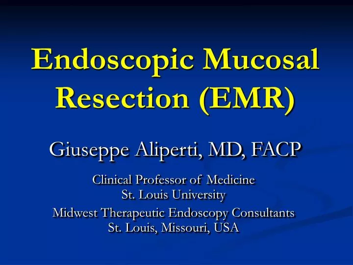
Endoscopic Mucosal Resection (EMR). Giuseppe Aliperti, MD, FACP. Clinical Professor of Medicine St. Louis University Midwest Therapeutic Endoscopy Consultants St. Louis, Missouri, USA . Endoscopic Mucosal Resection (EMR).

E N D
Endoscopic Mucosal Resection (EMR) Giuseppe Aliperti, MD, FACP Clinical Professor of Medicine St. Louis University Midwest Therapeutic Endoscopy Consultants St. Louis, Missouri, USA
Endoscopic Mucosal Resection (EMR) Major advance in minimally invasive surgery of the GI tract based on the following realities: • Endoscopy provides visualization/access to GI mucosa, where most cancers originate • Resection/retrieval of tissue allows pathologic examination (as opposed to ablation) First perfected in Japan for resection of superficial gastric cancer, very high in that Country unlike the West, where colon cancer (arising in polyps) is much more common
Endoscopic Mucosal Resection (EMR) • Most gastric cancers begin in slightly elevated, flat, or slightly depressed mucosal dysplastic lesions, difficult to grasp with a simple wire snare. • Most mucosal polyps, by projecting into the lumen, are easy to grasp with wire snares at the polyp base for resection with electrocautery
Endoscopic Mucosal Resection (EMR) Japanese endoscopists have adapted and perfected methods to raise the neoplastic mucosal area in order to allow snaring or dissection with sharp-tipped instruments Most use fluid injection into the submucosal layer to elevate the mucosa and allow it to be grasped with the snare Some use specially fitted scope caps that lift the lesion by suction
Endoscopic Mucosal Resection (EMR) The success of EMR in the stomach prompted endoscopists to expand the method to the: • Esophagus, where early cancer and premalignant dysplasia also tends to be non-polypoid and flat • Colon, where some neoplastic lesions are flat or sessile • Duodenum • Major papilla
EMR: techniques Special caps designed to fit the tip of the endoscope allow endoscopic suction to lift the mucosa to be ensnared Removal of large lesions in 1 piece (en bloc) is achieved with electrocautery wire-knives of different shapes that dissect around and under the diseased mucosa
High-Grade Dysplasia (HGD), Superficial Cancer in Barrett's Esophagus • Incresed detection rates with screening/surveillance • Difficult area in clinical decision making – HGD difficult to distinguish histologically from invasive adenocarcinoma intra-(T1m) or sub-mucosal (T1sm) • In specimens of esophagectomy for HGD >30% harbor invasive cancer • Esophagectomy has a high morbidity/mortality (2-7% in expert centers, 20% in others – age/comorbidities in older patients where HGD is more frequent)
Endoscopic Ablative Therapies (non-resective) methods based on rates of lymph node metastasis: absent in HGD, very low in confined invasive cancers 1-2-3 • photodynamic therapy • argon plasma coagulation Both successful4-5, but cannot assure: • Confinement to the mucosa – T1m vs. T1sm • Removal of the entire lesion - there is no specimen EUS accuracy, excellent for invasion beyond submucosa and lymph node detection - is only 75-85% in distinguishing between mucosal [T1m] and submucosal [T1sm] disease
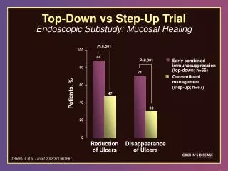
EMR for Staging in Barrett's Esophagus:General Concepts • EMR allows removal of large pathology specimens extending into the mid-submucosa, a big staging advantage over ablative methods 6 • Depth of cancer invasion can be made with great accuracy • Disease confined to mucosa with clear margins is considered cured • Patients with submucosal invasion (T1sm) are usually referred for surgery/chemo Ell et al Gastroenterol 2000;118:670-677
EMR for Staging in Barrett's Esophagus EMR performed to stage patients with HGD/T1m (confined to mucosa) CA by EUS 7 48 pts, 8 to surgery for submucosal (T1sm) disease by EUS: 7/8 T1sm, one T1m, overstaged 40 with T1m by EUS: 25 had HGD, 15 had adenoCA - All underwent EMR for definitive staging (no complications) EMR changed the staging in 30%: • 19/25 with apparent HGD by EUS were T1m1, 4/25 T1m2, 2/25 T1m3 • 6/15 with T1m CA were T1sm (submucosal invasion) instead and were sent to surgery additional therapy required for residual/recurrent HGD, CA Lightdale et al Gastrointest Endosc. 2004;59:AB90
Post-EMR Endoscopic Surveillance 240 EMR 1996-2003 8 • accurate assessment of invasion depth • neoplastic lesions not resected completely in most, required additional biopsies and f-u 72 EMR compared with 66 surgery for T1m/T1sm CA EMR patients older, smaller tumors, less LN mets 9 • EMR: fewer complications, shorter hospital stay • EMR: higher risk of tumor recurrence. Lewis et al, Mayo, GIE 2004;59:AB101 Bhave et al, MGH/UCH, GIE 2004;59:AB254
Post-EMR Endoscopic Surveillance 79 patients, 38 with HGD/T1m CA 10 • EMR alone satisfactory in 60% pts; T1sm invasion in 18% required surgery/chemo • Complete resection, clear margins not always possible EMR in 38 pts with HGD/T1m 11 • changed pretreatment diagnosis in 10 (26%) • negligible complications • careful endoscopic surveillance was recommended Ponchon et al, France, GIE 2004;59:AB255 Conio et al, Italy, GIE 2004;59:AB253
EMR for Barrett's Esophagus: Conclusions EMR performed for focal lesions or for complete removal of short-segment Barrett's EMR good for superficial cancer treatment, low complications Impossible to determine clear margins in piecemeal resections After EMR, careful follow-up is required for residual or metachronous disease
Circumferential EMR in Barrett's Esophagus All Barrett's tissue removed with special stiff monofilament snare in 22 patients with HGD/IMCA 12 • Tissue removed piecemeal, median length 3cm (1.2-10) • Four had residual Barrett's epithelium (1 beneath new squamous epithelium), 6 developed strictures responding to dilation Circumferential piecemeal EMR with modified multiple variceal band ligator, then multiple snare resections without removing scope in between 13 • greater efficiency then cap-assisted EMR for larger resections Seewald et al, Germany, GIE 2004;59:AB101 Soehendra, Germany, DDW 2004
EMR Combined With Ablation Therapy Used to treat residual neoplastic tissue after EMR Photodynamic therapy (PDT) with laser after light-sensitizing drugs successful method of ablation after EMR 14 Combination of EMR and PDT 15 • local remission in 26/28 pts, mean f-u 15.2 mo • local recurrence common, further treated with EMR+PDT Combination of EMR and PDT 16 • local remission in 18 of 22 pts over 10.6 months • no complications Pacifico et al, Clin Gastr Hepatol. 2003;1:252-257 Peters et al, The Netherlands GIE 2004;59:AB251 Haringsma et al, The Netherlands GIE 2004;59:AB252
EMR Combined With Ablation Therapy Combination EMR/PDT for focal neoplastic lesions followed with thermal ablation by APC 17 • Complete eradication of all of the Barrett's and all neoplastic tissue was achieved in 85/88 patients, mean f-u 30 months; 2 patients died of progressive disease • Complications included 1 bleed after EMR, 20 symptomatic strictures, 10 "sunburns," 2 episodes of atrial fibrillation after PDT Rahmani et al (IU) GIE 2004;59:AB250
EMR for Other Esophageal Tumors EMR for SCCA of esophagus is widely used in Japan, less in the West, where early detection is uncommon. EMR on 39 pts with early SCCA 18 • 10 pts CA in situ, 19 T1m, 10 T1sm (inoperable) • 9/10 with CA in situ, 19/19 patients with T1m with complete remission at mean f-u 29.7 mo • 3 minor bleeding, 3 mild strictures EMR (band ligation then snare) for submucosal benign tumors <3cm 19 • 17 pts: 11 granular cell tumors, 3 leiomyomas, 1 lipoma, 2 stromal • 7 bleeding, no transfusions (1 to surgery for residual) Pech et al, Germany GIE 2004;59:AB256 Wehrmann et al, Germany GIE 2004;59:AB241
Endoscopic Sub-mucosal Dissection Variation of EMR in which large resections are performed for accurate determination of margins and depth • Hyaluronic acid is injected to maintain long-lasting submucosal elevation • Dissection is performed with different shape devices through the endoscope operating channel • Experts are Dr. Haruhiro Inoue20 who uses a triangle-tipped knife Dr. Hiroyuki Ono21 who uses an insulated-tip knife • Dissections are lengthy, sometimes taking hours, and have significant perforation rates of 5-10%, most are managed endoscopically with clips Inoue, Japan, DDW 2004 Ono, Japan, DDW 2004
Endoscopic Sub-mucosal Dissection Adjustable thin-wire snare or a flex-knife 22 • 9 pts with GE-junction, cardia tumors • mean lesion size 20.8 mm, mean specimen size 35.2 mm • no significant bleeding, 1 stricture responding to dilation • 1 pt with submucosal invasion, all others without recurrence at mean f-u 8 mo. Yahagi et al, Japan, GIE 2004;59:AB171
Endoscopic Sub-mucosal Dissection Hook knife in left-sided colonic lesions 23 • 14 pts. mean diameter of 32.7 mm. En bloc resection in 10/14 (71%) pts • 1 microperforation with a 60mm flat rectal adenoma, managed with endoscopic clips and antibiotics Hotta et al, Japan GIE 2004;59:AB273
Ampullectomy: Protocol Removal of adenomas of the major papilla24 • Patients underwent biopsy, snare polypectomy, dual sphincterotomy with dual stent placement • Nd:YAG laser ablation or Argon Plasma Coagulation were used if the resection was felt to be incomplete • Endoscopy repeated at 4 to 8 weeks for stent exchange/removal with further ablation/biopsies until complete resection accomplished • Patients then followed with endoscopy/biopsies every 6 months and, if negative for recurrence, yearly Husain, Sawhney, Aliperti GIE 2001;53(5):3402
Ampullectomy: Results • Twelve pts 72 + 2.3 y (r 59-83) with jaundice/abnormal LFTs (58.2%), biliary colic (16.6%), Fe-def. anemia (16.6%) • Lesions between 2 and 5 cm (mean 2.5 cm) and normal cholangiography, except for dilatation • Median of 3 endoscopic sessions (range 2-4 ) for complete adenoma ablation • 50 % required Nd:YAG laser or APC Husain, Sawhney, Aliperti GIE 2001;53(5):3402
Ampullectomy: Results • At 40 mo (r 6-84 mo) recurrent adenomas in 6 (50%). Two with foci of adenoCA had Whipple; 4 had repeat endoscopic resection; disease free at 12, 21, 48 and 84 mo • Six (50%) patients had no disease recurrence. Two died of unrelated causes at 33 and 48 mo. The other four remain disease-free at 6, 22, 37 and 48 mo • On multivariate analysis: age, size, use of laser/APC not different between pts with and without recurrence • One pt developed cholangitis following stent occlusion and was successfully treated with antibiotics and stent exchange • No other endoscopy-related complications Husain, Sawhney, Aliperti GIE 2001;53(5):3402
Ampullectomy: Conclusions For patients with ampullary adenomas who are poor surgical candidates or refuse surgery: • Endoscopic resection with periodic surveillance is a safe and effective alternative • After endoscopic resection, age, size of adenoma and use of thermal therapy are not reliable predictors of recurrence Duodenal wall extension Adenoma before resection After mucosectomy, during laser ablation During healing, after stent removal
EMR of Choledochal Cyst • Not reported, so far… • Three cases, presenting with intermittant biliary obstruction (pain, LFTs) • Complete resection, no complications Aliperti, Unpublished
Conclusions • EMR is now firmly established as an important minimally invasive therapy for the treatment of mucosal cancer and premalignant lesions of the gastrointestinal tract. • The techniques for EMR are still in evolution. However, current methods involving submucosal fluid injection followed by electrocautery excision, or cap-assisted EMR using endoscopic suction to achieve mucosal lifting, have been highly safe and effective. • Endoscopic submucosal dissection allows large en bloc resections of larger superficial tumors, and this method has been proven safe and effective in expert hands.