ENDOSCOPIC MUCOSAL RESECTION
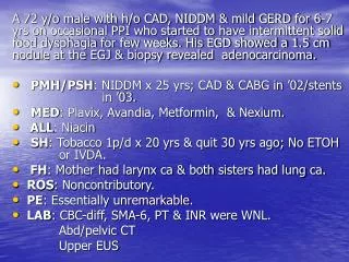
A 72 y/o male with h/o CAD, NIDDM & mild GERD for 6-7 yrs on occasional PPI who started to have intermittent solid food dysphagia for few weeks. His EGD showed a 1.5 cm nodule at the EGJ & biopsy revealed adenocarcinoma. PMH/PSH : NIDDM x 25 yrs; CAD & CABG in ’02/stents in ’03.

ENDOSCOPIC MUCOSAL RESECTION
E N D
Presentation Transcript
A 72 y/o male with h/o CAD, NIDDM & mild GERD for 6-7 yrs on occasional PPI who started to have intermittent solid food dysphagia for few weeks. His EGD showed a 1.5 cm nodule at the EGJ & biopsy revealed adenocarcinoma. PMH/PSH: NIDDM x 25 yrs; CAD & CABG in ’02/stents in ’03. MED: Plavix, Avandia, Metformin, & Nexium. ALL: Niacin SH: Tobacco 1p/d x 20 yrs & quit 30 yrs ago; No ETOH or IVDA. FH: Mother had larynx ca & both sisters had lung ca. ROS: Noncontributory. PE: Essentially unremarkable. LAB: CBC-diff, SMA-6, PT & INR were WNL. Abd/pelvic CT Upper EUS FH
A 77 y/o female with a COPD who has been seen by TMH/BCM liver team with an obstructive jaundice in Jan ’00 and referred for a surgical consult. Denied fever, wt loss, pruritis, GIB. • MED: Cipro, Actigall • PMH: COPD for 10-12 yrs. • SH: Tobacco 1 p/d for 50yrs, no ETOH or IVDA • FH & ROS: Noncontributory • PE: VSS, icteric & chest increased AP diamater, prolonged expirium, hepatomegaly. • LAB: WBC 11, H/H 14.6/41, Pl 171& lyts, FBS, PT, INR were all WNL. Alk.phos. 378, bil. 6.8 & ALT/AST 147/128. Abd US& abd/pelvic CT ERCP
ENDOSCOPIC PAPILLECTOMY in a 77 y/o female with adenoma 11/9/2000 5/12/2005
EMR in a 64 y/o male with 7 cm rectal villousadenoma 9/15/2000 4/21/2001 11/1/2001 6/17/2005
ENDOSCOPICMUCOSAL RESECTION (EMR) EMR first proliferated in Japan. The EMR combines the therapeuticpower ofendoscopic procedure with the diagnostic power of pathology exam of resected neoplastic lesion in selected cases. Saline assisted or cap-assisted EMR have been safe and effective for GI mucosal cancers. EMR using electrocautery knives has shown safe & effective for submucosal lesions in expert hands. Gastrointest Endosc 59: 171 & 273, 2004. Endoscopy 38: 521, 2005
ENDOSCOPIC PAPILLECTOMY (EP) • Ampullary adenomas can be removed by combination of EP & APC in selected cases after prophylactic pancreatic and sometimes biliary stenting. • These lesions can progress through an adenoma-carcinoma sequences. • Complications of EP occurred in about 20% such as bleeding, perforation & pancreatitis. • Depending upon the size & path of the lesion, appropriate post-EP surveillance is needed. Gastrointest Endosc 62: 367 & 551, 2005. Current Opinion in Gastroenterol 20: 40, 2004. Gut 53: 381,2004.