CELL STRUCTURES
CELL STRUCTURES Sources : Campbell, N. 1993 Biology Third Edition. Benjamin Cummings Publishing. United States. Wallace, S. 1993 Biology The Cytoskeleton The cytoskeleton (high voltage transmission electron microscope)

CELL STRUCTURES
E N D
Presentation Transcript
CELL STRUCTURES Sources: Campbell, N. 1993 Biology Third Edition. Benjamin Cummings Publishing. United States. Wallace, S. 1993 Biology
The Cytoskeleton • The cytoskeleton (high voltage transmission electron microscope) • Microfilaments (very fine; 6 nm in diameter). mostly made up of protein actin. Have a variety of roles depending on the type of proteins they are associated with: • one is to provide support to the plasma membrane • form supportive network of microvilli (intestinal wall) • also involved in the pinching of the cell membrane during cell division
Intermediate filaments (diameter 7 nm): Role is poorly understood. • Found in epithelial cells, nerve cells, muscle cell fibers and blood cells. • Made up of the protein keratin. • Microtubules (diameter 22 nm): • Formed from the globular protein tubulin • form cilia, flagella, centrioles, basal bodies • Important in cell division where they take a spindle form.
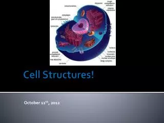
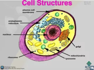
The Endoplasmic Reticulum (ER) Endoplasmic = inside the cytoplasm; Reticulum = network • Function • Plays a role in the cell’s synthetic ability: newly synthesized proteins, lipids and carbohydrates are transported within the endoplasmic reticulum’s lumen.
The ER ... • Structure: • The ER consists of membranous tubules and sacs called cisternae. Membranes separate the cytosol from the internal compartment of the lumen • Have the same phospholipid bilayer seen in the plasma membrane • The ER membrane is continuous with the outer of the two membranes of the nuclear envelope
The Rough Endoplasmic reticulum (RER) • Occurs in cells that manufacture proteins for secretion outside of the cell (i.e digestive enzymes and hormones) • The Smooth Endoplasmic reticulum (SER) • Abounds in cells that synthesize secrete, and store carbohydrates, steroids, lipids and other non-protein products. • Found in testis cells; oil glands; some hormone producing gland cells; intestinal cells, where they collect products of lipid digestion; in liver cells the SER is associated with glycogen, the animal storage carbohydrate (starch).
Ribosomes • Structure: Large molecular structures consisting of ribosomal rRNA sub-units and proteins. • Consists of 2 sub-units (large and small) which join together to form a functional ribosome only when they attach to mRNA • Ribosomes are constructed in the nucleolus from RNA • Are too large to be considered molecules. The ribsomal sub-units in prokaryotes are smaller and differ chemically.
Ribosomes... • Function: Where the cell assembles proteins following genetic (DNA) instructions. Ribosomes translate message from mRNA • There are two forms: • Polyribosomes or polysomes: Free • Bound ribosomes: Attached to the ER
The Golgi Apparatus Sometimes called Dyctiosomes in plant cells • Structure (see diagram) • Component of the endomembrane system • Made up of flattened bag-like sacs called cisternae. Found near the nucleus. (cis phase = forming face; trans face = maturing phase) • Function • Manufacturing, warehousing, sorting, and shipping center of materials in the cell
Mitochondria • Structure (see diagram) • Have double membranes • Have their own circular DNA and ribosomes (make their own proteins) • Smaller than chloroplasts • develop from pre-existing mitochondria
Mitochondria... • Function • ATP (adenosine tri-phosphate) generating molecules
Peroxisomes (Microbodies) • Structure: • Sacs bound by a single membrane • Function: (see transparency) • organelles found in nearly all eukaryotic cells that contain specialized enzymes for specific metabolic activities • All contain peroxide-producing oxidases (enzymes) that transfer H from various substrates to O producing H2O2