Download
1 / 51
510 likes | 616 Views
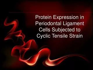
Protein Expression in Periodontal Ligament Cells Subjected to Cyclic Tensile Strain. Content Outline. Introduction Materials & Methods Results Discussion Conclusion. Introduction. Background.

E N D
Protein Expression in Periodontal Ligament Cells Subjected to Cyclic Tensile Strain
Content Outline • Introduction • Materials & Methods • Results • Discussion • Conclusion
Background • Orthodontics is a discipline specialised in the study and treatment of malocclusions, with the purpose of improving appearances and correcting deranged and unstable occlusion http://www.harrisongarrisondentistry.com/beforeafter1revised_edited-1.jpg After Before
Background • Demand for orthodontics remains high and with it, an increasing interest in the mechanisms of tooth movement.
Background • Mechanical forces applied on teeth generates tensile and compressive forces at the PDL interface within the alveolar bone. Orthodontic force Area under tension Area under compression
Background • Corresponding bone deposition and resorption then occurs. • The tooth thus migrates to its desired spatial position. Orthodontic force Bone deposition Bone resorption
Background • The focus of academic interest has been centered on the metabolic profile at the PDL interface. • Studies have been done to identify cytokines responsible for inducing osteoblastic and osteoclastic activities at the cellular and molecular level.
Objectives of Experiment • To investigate the proteins expressed by different genes of interest, as found in the study conducted by Pinkerton et al (2008), when human PDL cells are subjected to uniform radial & circumferential strain. Pinkerton et al (2008)
Preparation of Human PDL Cells • Prior to extraction, localised subgingival scaling was done. • Pericision under local anaesthesia was carried out. • Teeth were then extracted.
Preparation of Human PDL Cells • Extracted teeth were immersed in Falcon tubes containing pre-incubated culture medium and transported to the laboratory.
Preparation of Human PDL Cells • PDL from the middle 1/3 of the root was removed and plated onto 35mm petri-dishes. • Culture medium was then added.
Preparation of Human PDL Cells • Cells were placed in a CO2 incubator with a humidified atmosphere of 5.1% CO2 and 95% air at 37.0°C.
Preparation of Human PDL Cells • Media was changed at weekly intervals. • Any contamination and outgrowth of cells from the tissue explants were examined under light microscope.
Preparation of Human PDL Cells • Once confluence was reached by primary explanted cells (P0), they were trypsinized and cultured in a 100mm Petri dish. • This 1st passage of cells was labeled P1. non-confluence 100% confluency
Preparation of Human PDL Cells • From P1, whenever confluence was attained, cells were trypsinized and serially transferred to 75 cm2 (P2) and 175 cm2 (P3) tissue culture flasks. • The 4th passage of cells (P4) was used for this experiment. P1 P2 P3 & P4
Application of Tensile Strain to PDL Cells • P4 cells were subcultured into the 6-well, flexi-bottomed BioFlex® culture plates. BioFlex® culture plate
Application of Tensile Strain to PDL Cells • Each BioFlex® culture plate was placed on a Loading Station™ which consist of 6 loading posts designed to fit onto the flexi-bottom of the BioFlex® culture plate. Loading posts BioFlex® culture plate Loading Station™
Loading Station™ 4 BioFlex® culture plates VACUUM GENERATOR Flexercell FX-4000 Strain Unit Application of Tensile Strain to PDL Cells • The Loading Station™ is then connected to a vacuum generator regulated by the Flexercell FX-4000 Strain Unit, and placed in a CO2 incubator. CO2 INCUBATOR
Application of Tensile Strain to PDL Cells • When vacuum is applied to the BioFlex® culture plates, the rubber membrane carrying the PDL cells will deform across the loading post face, creating uniform equibiaxial strain. PDL cells BioFlex® culture plate Equibiaxial strain REST Rubber membrane Loading Post VACUUM SIDE VIEW TOP VIEW
Application of Tensile Strain to PDL Cells • The cells were subjected to an intermittent deformation of 12% for 5 sec every 60 sec with the Flexercell FX-4000 Strain Unit. 12%
6 HOURS • 12 HOURS • 24 HOURS Application of Tensile Strain to PDL Cells • Four plates (2 experimental and 2 control) were allocated to each of the 3 time intervals: 6, 12, and 24 hrs. Experimental : Control :
Loading Station™ 4 BioFlex® culture plates VACUUM GENERATOR Flexercell FX-4000 Strain Unit Collecting and Preparing the Supernatant • The FlexercellStrain Unit was paused temporarily to allow BioFlex® culture plate removal at the end of each time interval. 12 HOURS 24 HOURS 6 HOURS CO2 INCUBATOR
Collecting and Preparing the Supernatant • Culture media is then removed from the wells and stored at -20C. -20C
Assaying the Proteins • Samples collected were sent for quantitative assaying by ELISA for the following cytokines:
Problems Encountered • Contamination 5x magnification 10x magnification
Problems Encountered • Unpredictable growth rate of cells • Failure to reach confluency • Failure of cells to attach to Bioflex® wells • Flexercell software • Not configured appropriately • Delay resulted in differentiation of cells
Mechanism of Cellular Response • The mechanism by which human PDL cells produce cytokines when subjected to mechanical strain has not been proven. • Some postulate that mechanical strain may lead to deformation of blood vessels supplying the PDL cells, causing local hypoxia, which induces the PDL to produce chemical mediators as an adaptive response.
12% Equibiaxial Strain • Equibiaxial strain more closely represent deformation experienced by PDL cells during orthodontic tooth movement • 12% selected based on numerical data derived from a finite element model: • Maximal PDL strains for horizontal displacements of human maxillary central incisor under physiological loading conditions within 8-25%, dependent on apico-crestal position • 12% correlates well with strain values predicted at mid-root
Cytokines • Cytokines are low-molecular weight proteins produced by cells, which regulate or modify action of other cells in an autocrine or paracrine manner • Autocrine: acting on cell of origin • Paracrine: acting on adjacent cells • Cytokines include interleukins (ILs), growth factors etc
Interleukins • IL-1 family are pro-inflammatory cytokines, having been shown to stimulate bone resorption • Also seen in periodontal diseases • IL-6, like IL-1, stimulates bone resorption and induces osteoclast proliferation • IL-1 has been shown to induce IL-6 production
Interleukins • IL-7 is a potent osteoclastogenic factor capable of inducing bone loss when administered in vivo • Elevated levels have been associated with Rheumatoid Arthritis (RA), a condition causing systemic bone loss • IL-8 stimulates osteoclastogenesis • GCF of persons with periodontal disease contain higher levels of IL-8
Interleukins • IL-11 enhances bone resorption by promoting osteoclastogenesis and by suppressing the activity of osteoblasts • Can stimulate bone resorption, but can also work with BMP-2 to induce osteoblastic differentiation – bone formation • IL-12A inhibits osteoclast formation and bone resorption
Other Cytokines • RANKL (Receptor Activator of Nuclear Factor Kappa B Ligand) • Expressed by activated cells during periods of mechano-compression • Bind to RANK membrane receptors to activate osteoclasts, causing bone resorption • Cell – cell signalling by RANKL is essential for the induction of osteoclast differentiation
Other Cytokines • OPG (Osteoprotegerin) • Decoy receptor for RANKL • Inhibition of osteoclast differentiation and resorptive function • Stimulation of osteoclast apoptosis • Suppression of bone resorption
IL-1A & IL-1F7 • IL-6 • IL-7 Summary of Role of Cytokines • IL-8 • IL-11 • RANKL • OPG • IL-12A BONE RESORPTION BONE DEPOSITION
Pinkerton et al (2008) • Human PDL cells were strained with cyclic deformation of 12% for 6, 12 and 24 hours • Differential expression of 79 cytokine and growth factor genes in the form of mRNA was quantified using real-time PCR arrays
IL-1A & IL-1F7 • IL-6 • IL-7 Pinkerton et al (2008) • IL-8 • IL-11 • RANKL • OPG • IL-12A • These were the findings: DOWN-REGULATED GENES UP-REGULATED GENES NO SIGNIFICANT CHANGE BONE DEPOSITION BONE RESORPTION
Gene Expression vs. Protein Expression • Not all expressed genes (in the form of mRNA) are translated into proteins. • Up-regulation in gene expression does not represent up-regulation in cytokine or growth factor production, and vice versa.
Limitations of Our Study • PDL cells in 2-dimensional substrate • Not an accurate real-life representation • Real-life PDL cells exist in a 3-dimensional matrix and experience mechanical forces in multiple planes • Biologic activity of proteins not tested • Proteins may be expressed but do not exert any influence in cellular signalling
Limitations of Our Study • Small number of gene products analysed • Cytokine biology is highly complex • Effects of each cytokine is wide-ranging (pleiotropy), with overlapping biologic activities (redundancy) • Cytokines are analysed with the assumption that they act singly rather than in unison • Other protein factors associated with these cytokines function were not taken into account • E.g. Bone Morphogenic Protein-2 for IL-11
Limitations of Our Study • One source of PDL cells • Studies have shown that there is individual variant in tissue response during orthodontic treatment • Amount of cytokine expression may vary in different PDL cells obtained from different patients • Use of intermittent 12% strain • Continuous strain would better mimic effect of orthodontic appliance on PDL cells
Concluding Remarks • Orthodontic tooth movement relies on cellular signalling molecules produced by PDL cells subjected to mechanical strain, with resultant bone remodelling which allows tooth migration in the direction of the orthodontic force. • A large array of cytokines and growth factors have been identified in association with mechanical deformation of PDL cells, both in in vitro and in vivo models.
Concluding Remarks • The knowledge of the exact role each of these biological molecules as well as the interaction between them, plus their expression in response to different forms, magnitude and frequency of strain experienced by the PDL cells remain incomplete. • Intepretation of results from in vitro models such as our experiment should be correlated with in vivo data obtained from animal models, but both types of models have their limitations that must be accounted for.
Concluding Remarks • Future investigations should be directed towards studying PDL cells in an artifical 3-dimensional matrix created from collagenous gel matrix, which will resemble conditions in vivo. • Biologic activity of protein products collected should also be looked into, using the appropriate bioassays. • Additional studies could also focus on the cytokine biology involved in terms of complex interacting networks, with individual mediators acting in unison rather than singly
Acknowledgements • Professor Murray Clyde Meikle • Dr Loh Hwee Hiang • Dr Vinoth Kumar • NUS Academic Research Fund Grant