Download
1 / 65
2.01k likes | 6.95k Views
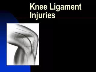
Periodontal ligament. Contents. Introduction Definition Development of PDL Formation of PDL Structure Collagen fibers Formation of collagen Functions Blood supply Nerve supply Lymphatics. Age changes Clinical considerations. Introduction.

E N D
Contents • Introduction • Definition • Development of PDL • Formation of PDL • Structure • Collagen fibers • Formation of collagen • Functions • Blood supply • Nerve supply • Lymphatics
Age changes • Clinical considerations
Introduction PDL is the soft, richly vascular and cellular connective tissue which surrounds the roots of teeth and joins the root cementum with the socket wall.
Definitions • Is the connective tissue that surrounds the root of the tooth and connects it with the alveolar bone. • Together with cementum, alveolar bone and lamina propria of gingiva, PDL forms the tissue which supports the tooth in the jaw. These supporting structures are often referred to collectively as PERIODONTIUM
Over the years it has been described by a number of terms like • Desmodont • Gomphosis • Pericementum • Dental periosteum • Alveolo dental ligament • Periodontal membrane However because it is a complex soft connective tissue providing continuity between two mineralized connective tissues, the term PDL appears to be more appropriate.
Extent and shape • In the coronal direction it is continuous with lamina propria of gingiva & is demarcated by the alveolar crest fibers. At the root apex it merges with the dental pulp.
PDL has the shape of an hour glass and is narrowest at the mid root level. It ranges in width from 0.15-0.38mm. avg width 0.21mm ----11-16 yrs 0.18mm ---- 32-52 yrs 0.15mm ---- 51-67 yrs Width also varies according to functional state of the tissues time of eruption ---- 0.1- 0.5mm at function ---- 0.2-0.35mm hypo function ---- 0.1-0.15mm
Development of PDL The periodontal tissues that support a tooth in the jaw are derived from dental follicle which in turn originates from dental papilla. It has also been proposed that the mesenchyme deriving periodontium may have two differentiation compartments a. Alveolar clade --- Fibroblasts & osteoblasts b. Cementum clade --- fibroblasts & cementoblasts
Formation of PDL occurs after the cells of HERS have separated, forming the strands epithelial rests of malassez. This separation permits the cells of dental follicle to migrate to the external surface of the newly formed root dentine. The fibers of PDL become embedded in newly developed cementum & alveolar bone and as the tooth erupts are oriented in characteristic fashion.
Organisation of Periodontal Ligament Tooth bud is formed in a crypt of bone. The collagen fibers produced by the fibroblasts in the loose connective tissue around the tooth bud are embedded into the newly formed cementum immediately apical to the CEJn. The true PDL fibres (Principal fibers) develop in conjunction with tooth eruption.
Formation of PDL • Collagen fibers are embedded into newly formed cementum apical to CEJ. They orient towards the coronal portion of the bony crypt which later form the dentogingival , dentoperiosteal & transseptal fiber groups. • The true PDL fibers the principal fibers develop in conjunction with the eruption of the tooth. • First fibers are identified entering the most marginal portion of the alveolar bone.
Later more apically positioned bundles of oriented collagen fibers are seen. • The orientation of collagen fiber bundles alter continuously . When tooth has reached contact in occlusion and is functioning properly the fibers of PDL associate into groups of well oriented dento alveolar collagen fibers which undergo constant remodeling.
Later, more apically positioned bundles of oriented collagen fibres are seen. The orientation of the collagen fiber bundles gets altered continously during tooth eruption and stabilises once the tooth reaches in occlusion.
Development of Principalfibers First, small, fine, brush-like fibrils are detected arising from the root cementum and projecting into the PDL space. The surface of the bone is covered by osteoblasts. Later the number and thickness of fibers entering the bone increase. The fibers originating from the cementum are still short while those entering the bone gradually become longer. The fibers originating from the cementum subsequently increase in length and thickness and fuse in the periodontal ligament space with the fibers originating from the alveolar bone.
Terminal portions of principal fibers that insert into cementum and bone are termed as Sharpey’s fibers. The principal fibers embedded in the cementum have a small diameter but are more numerous than those embedded in the alveolar bone proper. In addition to these fiber types, small collagen fibers associated with larger principal fibers have been called as “Indifferent fiber plexus of Shefforfold”. Other features of developing collagen fibers: • Bundling mechanism • Cushion hammock ligament • Pulp limiting membrane • Crimping effect
Structure • Cellular elements a. connective tissue cells SyntheticResorptive Fibroblasts Fibroblasts Cementoblasts Cementoclasts Osteoblasts Osteoclasts b. Epithelial cell rests c. Immune system cells d. Neuro vascular elements • Periodontal fibers • Ground substance
a.Connective tissue cells • Importantly fibroblasts, cementoblasts & osteoblasts • Fibroblasts are the most common cells in PDL and appear as ovoid or elongated cells oriented along the principal fibers, exhibiting pseudopodia like processes. • These cells synthesize collagen and also have the ability to phagocytose old collagen and degrade them by enzyme hydrolysis.
Thus collagen turn over appears to be regulated by fibroblasts in a process of intra cellular degradation. • Phenotypically distinct & functionally different sub populations of fibroblasts exist in adult PDL. • They may have different functions like secretion of different collagen types & production of collagenase.
Bone & Cementum cells • Although technically situated within the PDL, bone and cementum cells are properly associated with the hard tissues they form. • Osteoblasts line the bone surface of the ligament and may be either functional or resting, depending on the functional state of the ligament • Osteoclasts may be found against the bone where resorption is taking place. • Cemetoblasts are responsible for formation of cellular cementum.
cementoblasts osteoblasts
b.Epithelial rests of Malassez (Malassez 1884) • They are remnants of HERS and are formed close to cementum and are most numerous in the apical area & cervical area. • They form a lattice work and appear as either isolated cluster of cells or interlacing strands. They diminish in number with age and may undergo calcification to form cementicles. • Surrounded by basal lamina & inter connected by hemidesmosomes. Contain kerationocyte growth factors. Can proliferate and participate in formation of peri apical cysts and lateral root cysts.
Fig. shows the presence of clusters of epithelial cells (ER) in the periodontal ligament. These cells, called the epithelial cell rests of Mallassez, represent remnants of the Hertwig's epithelial root sheath. The epithelial cell rests are situated in the periodontal ligament at a distance of 15-75 μm from the cementum (C) on the root surface. A group of such epithelial cell rests is seen in a higher magnification.
c. Defense cells Include neutrophils, lymphocytes, macrophages,& eosinophils & mast cells. Mast cells: Small round or oval cell. Diameter 12-15µm. numerous cytoplasmic granules. Contain heparin & histamine. Role of heparin is not clear. Histamine plays a role in inflammatory reaction. Occasionally seen in healthy PDL. It may cause proliferation of endothelial & mesenchymal cells.
Macrophages • Predominantly located adjacent to blood vessels. • Has a nucleus , regular contour, horse shoe shape or kidney shape. • Dual role a. phagocytosing dead cells b. secreting growth factors that regulate the proliferation of adjacent fibroblasts.
Periodontal fibers The most important elements of PDL are the principal fibers which are collagenous. They are associated with abundant non collagenous proteins typically found in bone and cementum They are thought to contribute to the regulation of mineralization and to tissue cohesion at sites of increased bio mechanical strain.
Collagen • Is derived from the French word collagene to designate connective tissue constituents that produce glue. • The collagen molecule is rigid and resists stretching; therefore it is utilized in tissues where mechanical forces should be transmitted without loss. • The organization of collagen depends upon the specific functional requirements in various tissues.
It is a protein compose of amino acids. The amount of collagen in a tissue is determined by its hydroxyproline content. Collagen classes; a. Interstitial collagens ---- Type I,II,III b. Basement membrane type---- Type IV,VI,VII c. Short chain collagens---- Type IX,X Theprincipal fibers are composed mainly of type I where as reticular fibers type III.
Collagen biosynthesis occurs inside the fibroblasts to form tropocollagen molecules . • These aggregate into microfibrils that are packed together to form fibrils • Collagen fibers have a transverse striation with the characteristic periodicity of 64nm, this striation is caused by the overlapping arrangement of the tropocollagen molecules.
Fibrillogenisis • The procollagen molecules are assembled extracellularly in the form of typical banded fibril. The entire process of assembly is known as fibrillogenisis.
PRINCIPAL FIBERS OF PDL • Transseptal • Alveolar crest group • Horizontal • Oblique • Apical • Inter – radicular Principal fibers of PDL
1.Transseptal Group • Extend Interproximally over the alveolar bone crest and are embedded in the cementum of adjacent teeth. • Are a remarkably constant finding and are reconstructed even after destruction of the alveolar bone has occurred in periodontal disease. • Considered as belonging to the gingiva because they do not have osseous attachment.
PDL TRANS-SEPTAL FIBER GROUP TOOTH TOOTH TRANS-SEPTALin medio-distal plane GINGIVA Alveolarseptum
2.Alveolar Crest • Extend obliquely from the cementum just beneath the JE to the alveolar crest. • Fibers also run from the cementum over the alveolar crest and to the fibrous layer of the periodontium covering alveolar bone • The alveolar crest fiber prevent extrusion of tooth and resist lateral tooth movement
3. Horizontal Group • Extend at right angles to the long axis of tooth from cementum to the alveolar bone. 4. Oblique group • Largest group in the PDL extend from the cementum in a coronal directing obliquely to the bone. • Bear the brunt of vertical masticatory stresses and transform them into tension on the alveolar bone.
5. Apical Group • Radiate in a rather irregular fashion from the cementum to the bone at the apical region of the socket . • Do not occur on incompletely formed roots. 6. Inter Radicular Fibers • Fan out from cementum to the tooth in the furcation areas of multirooted teeth. Other well formed fiber bundles interdigitate at right angles or splay around.
Oxytalan fibers • Restricted to walls of blood vessels in humans • PDLfibers does not contain mature elastin but two immature forms are found oxytalan and eluanin. • Oxytalan fibers run parallel to the root surfaces in vertical direction and bend to attach to cementum in the cervical third of the root.
An elastic meshwork has been described in the PDL as being composed of many elastin lamellae with peripheral oxytalan & eluanin fibers • Functions • Regulate vascular flow • Role in tooth support • Facilitate fibroblast attachment and migration
GROUND SUBSTANCE • Fills the space between the fibres and cells • Consists of a biochemically complex, highly hydrated, semisolid gel. • Water content of 70% Glycasaminoglycans – hylaluronic acid, proteoglycans( versican , decorin ) Glycoproteins ---- fibronectin , laminin , vibronectin , tenascin
FUNCTIONS OF PDL - Physical - Formative and Remodeling - Nutritive - Sensory
Physical functions 1) Provision for a soft tissue ‘CASING’ to protect the vessels and nerves from injury to by mechanical forces . 2) Transmission of occlusal forces to the bone 3) Attachment of teeth to bone. 4) Maintenance of gingival tissues in their proper relationship to the teeth. 5) Resistance to impact of occlusal forces
Resistance to impact of occlusal forces Shock absorption 1) Tensional theory 2) Viscoelastic theory • Thixotrophic theory
TENSIONAL THEORY- ascribes the principle fibers the major responsibility in transmitting the forces to the bone. • VISCOELASTIC THEORY- displacement of tooth largely controlled by fluid movements with fibers having only secondary role
Thixotropic theory Behaviour of tooth with axial intrusive load was consistent with the properties of a thixotropic material . A thixotropic material is the one which can undergo gel/sol/geltransformation It has the following rheological characteristics. • An isothermal change in viscosity is brought about by pressure alone • The system undergoes a time dependent recovery.
Transmission of occlusion forces to bone • Arrangement is like suspension bridge or hammock. • The oblique fibers alter their wavy pattern and sustain the major part of the axial force