Download
1 / 20
210 likes | 830 Views
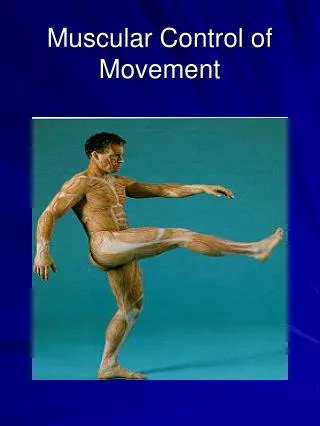
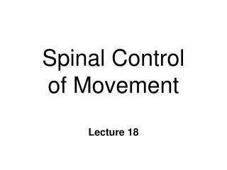
Spinal Control of Movement. Lesson 19. Anatomy. Ventral Spinal Cord Topographic organization Alpha motor neurons Spinal interneurons Striate muscle extrafusal fibers ~. Distal limbs. Proximal limbs. Alpha Motor Neurons. Or lower motor neurons (Class I) Cell body in ventral horn

E N D
Spinal Control of Movement Lesson 19
Anatomy • Ventral Spinal Cord • Topographic organization • Alpha motor neurons • Spinal interneurons • Striate muscle • extrafusal fibers ~ Distallimbs Proximallimbs
Alpha Motor Neurons • Or lower motor neurons (Class I) • Cell body in ventral horn • Emerge from ventral root • Innervate extrafusal fibers • Uninterrupted to muscle fibers • final common pathway • Only excitatory input to muscles Inhibition at spinal cord ~
Dorsal Extrafusal Fibers Alpha Motor neuron Ventral ACh
Input to Alpha Motor Neurons • 3 sources only 1. DRG neurons • sensory neurons (proprioception) • feedback from muscle spindles 2. Upper motor neurons • primarily from M1 3. Spinal interneurons • largest input (excitatory & inhibitory) • generation of motor programs ~
Inputs to Alpha Motor Neurons Sensory neurons Dorsal DRG Spinal interneurons Upper motor neurons - M1 Ventral
Neuromuscular Junction • Synapse between neuron & effector • Cholinergic (ACh) • nicotinic receptors • Motor end-plate • postsynaptic membrane • folds packed with receptors • increased surface area ~
Motor end-plate Terminal Button Muscle Fiber
Neuromuscular Organization • Motor Units • Single alpha motor neuron and all the muscle fibers that it innervates • 1:3 to 1:100 • fewer fibers finer control • Motor Pool • all alpha motor neurons that innervate a single muscle ~
Graded Control of Muscle Contraction • Highly reliable synapse 1 presynaptic AP 1 postsynaptic AP 1 twitch (contract/relax) • temporal summation • tension & sustained contraction • Recruitment • # motor units tension • order: smallest largest ~
Extrafusal Muscle Fibers • Striate muscle • Force for limb movements • flexion - closes joint • extension - opens joint • Contract or relax ~
Muscle Contraction • AP generated in muscle fiber (cell) • Ca++ released from internal stores • Muscle fiber contracts • continues while Ca++ & ATP available • Relaxation • Ca++ sequestered by active transport ~
Movement of Limbs • Flexors and extensors are ANTAGONISTIC • muscles and are reciprocally innervated • Limb flexion • flexors excited & extensors inhibited • Limb extension • extensors excited & flexors inhibited • Disynaptic inhibition ~
Dorsal + + - + Alpha Motor neurons Ventral Upper Motor Neurons +
Withdrawal Reflex • Flexion • remove limb from noxious stimulus • Polysynaptic reflex • sensory neuron • interneurons • motor neuron • 2 or more synapses • slower than monosynaptic ~
+ + - Polysynaptic withdrawal reflex + + + R
Central Pattern Generators • Half-center Model • alternating activity in flexor & extensor • Step-cycle has 2 phases • swing phase • foot off ground & flexing upward • stance phase • foot planted & leg extending • Each limb has own pattern generator ~
+ + + + + + + + Half-center Model Flexor a Tonic input a Extensor
Rhythmic Patterns: Sensory Feedback • Not necessary for locomotion • but slower, less coordinated • Stumble correction reaction • during swing phase • tactile stimulus on dorsal foot flexion • Reflex reversal • override during extension • flexion would cause collapse ~