Download
1 / 40
400 likes | 522 Views
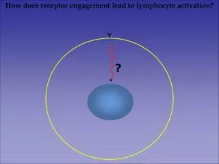
How does receptor engagement lead to lymphocyte activation?. Y. ?. Basic signal transduction. -signals are transmitted through changes in specific activity or location of proteins

E N D
How does receptor engagement lead to lymphocyte activation? Y ?
Basic signal transduction -signals are transmitted through changes in specific activity or location of proteins -these changes in protein function are mediated by binding small molecules (e.g. Ca++) or by covalent modifications (e.g. phosphorylation), and also by protein-protein interactions
PO4- Tyr Pro Pro Pro Interaction domains found in signaling proteins can modify function through changes in activity and/or localization. Domain Ligand Phosphotyrosine SH2 Proline-rich sequences SH3 Phosphatidylinositol Triphosphate (PIP3) PH
-P-P-P- Three types of tyrosine kinases are involved in antigen receptor signaling. Each has a conserved catalytic domain and protein interaction domains Src kinase family (e.g. Lck, Fyn, Lyn, Blk) Syk/ZAP-70 family Tec kinase family (e.g. Itk,Btk,Rlk) SH3 SH2 Catalytic domain
Tyrosine kinase activity is regulated by tyrosine phosphorylation and protein interaction domains Lck The Csk kinase phosphorylates the inhibitory tyrosine and the CD45 phosphatase can dephosphorylate this tyrosine. Src kinases autophosphorylate the activating tyrosine.
Activation loop phosphorylation alters the catalytic domain structure of Src kinases
Receptor clustering can signal the presence of bound ligand by juxtaposition of the cytoplasmic domains of the receptor. Association of kinase domains, present in many growth factor receptors, leads to kinase activation and the initiation of intracellular signaling.
Figure 6-3 Aggregation of receptors may lead to their localization in membrane rafts where Src kinases can initiate signaling
In some cases, receptor activity directly modifies transcription factor function - cytokine receptor signaling to STAT transcription factors
MAP kinase signal transduction pathway: Guanine nucleotide exchange factors (GEFs) activate small G-proteins to initiate a sequence of kinase phosphorylation events which culminate in transcription factor activation
Phosphatidylinositol signaling pathway: Phospholipase C activity generates second messengers, inositol trisphosphate (IP3) and diacylglycerol (DAG) which increase levels of intracellular calcium and activate Protein kinase C (PKC).
Adaptor proteins have no catalytic activity, but can provide binding sites which bring together effector and target proteins to promote efficient coupling of receptors to intracellular signaling pathways
Non-polymorphic components of the B and T lymphocyte antigen receptors mediate signaling
e e g d z z Immunoreceptor Tyrosine-based activation motifs (ITAMs) are present in multiple copies in the cytoplasmic domains of antigen receptors a b hz1 Q L Y N E L N L G R R E E - Y D V L hz2 G L Y N E L Q K D K M A E A Y S E I hz3 G L Y Q G L S T A T K D T - Y D A L hCD3g Q L Y Q P L K D R E D D Q - Y S H L hCD3e P D Y E P I R K G Q R D L - Y S G L hCD3d Q V Y Q P L R D R D D A Q - Y S H L mIga N L Y E G L N L D D C S M - Y E D I mIgb H T Y E G L N I D Q T A T - Y E D I
e e g d z z z z ITAM motifs were shown to be sufficient for signaling when expressed as chimeras, independent of the receptor CD8 a b
Membrane mobility and cytoskeletal function allows TCR’s that bind MHC-peptide to cluster at the T cell-APC interface.
Src family kinases are responsible for the tyrosine phosphorylation of ITAM motifs in antigen receptors
-P e e e e d d g g z z z z P- P- P- P- Receptor phosphorylation leads to recruitment and activation of the ZAP-70 tyrosine kinase a b a b Lck ZAP-70
Y PTKs Initiation of Antigen Receptor signaling depends upon non-receptor protein tyrosine kinases (PTKs)
Models of antigen receptor signal initiation Receptor-associated kinase aggregation leads to kinase activation and ITAM phosphorylation. 2. Receptor aggregation leads to co-localization with kinase and ITAM phosphorylation.
1. Receptor aggregation leads to co-localization of receptor and kinase. 2. Kinase phosphorylates receptor 1. Receptor aggregation leads to transphosphorylation and activation of receptor-associated kinase. 2. Kinase phosphorylates receptor Potential mechanisms regulating the initiation of TCR signaling by Src kinases Co-localization model Activation model -P P-
CD4 and CD8 co-receptors bind to Lck and help initiate signaling by delivering the kinase to the cytoplasmic domains of the T cell receptor following binding to MHC-peptide
Y PTKs ?
Protein tyrosine kinases use adaptor proteins to link antigen receptors to intracellular phosphatidylinositol (PI) and MAP kinase pathways Y PTKs adaptors PI pathway MAP kinase pathway
ZAP-70 phosphorylates two adaptor molecules: Linker for Activated T cells (LAT) and SH2 domain containing Leukocyte Protein (SLP-76)
R a s Ras G T P GDP Raf Y-PO 4 SOS MEK ERK The transmembrane adaptor LAT recruits SOS, a GTP exchange factor, to the membrane where it activates Ras and initiates the (ERK) MAP kinase pathway GRB2 LAT MEK
~P ~P ~P The LAT and SLP-76 adaptors co-localize PLCg and the Tec family kinase ITK. Activation of PLCg by ITK induces cleavage of PIP2 to DAG and IP3. DAG PLCg1 IP3 ITK LAT P~ SLP-76
P~ ~P P~ ~P LAT LAT SOS SOS GRB2 GRB2 GRB2 GRB2 Each SOS exchange factor can bind two Grb2 adaptor molecules Houtman, et al. Nature Structural Biology 13: 798-805, 2006
Mutation of the tyrosine phosphorylation sites in the carboxy-terminus of LAT that mediate Grb2 binding prevents LAT clustering following TCR stimulation
Signaling complex (signalsome) formation mediated by multivalent protein interactions
Regulation of gene expression through increased transcription factor activity NFAT: nuclear localization regulated by calcium-activated phosphatase AP-1: expression and activity regulated by MAPK pathways NFkB: nuclear localization regulated by degradation
The NFAT transcription factor is normally phosphorylated and sequestered in the cytosol. The PI pathway induces calcium influx which activates Calcineurin phosphatase activity and promotes nuclear translocation of the NFAT transcription factor.
Two different MAPK pathways are involved in lymphocyte activation
MAP kinase pathways increase AP-1 dependent transcription by increasing expression of the Fos subunit, and by increasing activity of the Jun subunit Fos -P -P
Stimulated antigen receptors utilize tyrosine kinases to induce intracellular signaling pathways leading to transcription factor activation and the induction of cell growth and differentiation. Y PTKs adaptors PI pathway MAP kinase pathways Interleukin-2, and effector functions
The NF-kB transcription factor is sequestered in the cytosol by IkB which is phosphorylated and degraded in response to receptor signals nucleus
Rawlings et al.Nature Reviews Immunology6, 799–812. 2006) Antigen receptor activation of NF-kB depends upon assembly of a CARMA - BCL10 - MALT - TRAF complex that leads to the ubiquitylationof IKKassociation of TAK, and IKK phosphorylation