Download
1 / 37
370 likes | 523 Views
Blood and Immunity. What is Blood?. Blood is a fluid tissue. It is important for the protection and survival of all our bodies cells. The average person has about 5 L of blood. Of this: 55% is fluid called plasma - mainly water, minor dissolved O2, CO2,

E N D
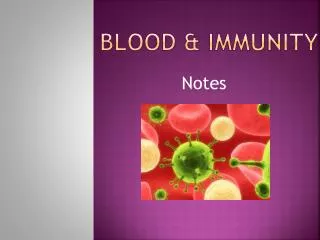
What is Blood? • Blood is a fluid tissue. It is important for the protection and survival of all our bodies cells. The average person has about 5 L of blood. Of this: • 55% is fluid called plasma - mainly water, minor dissolved O2, CO2, nutrients, waste, salts, hormones and vitamins • <1% is white blood cells and platelets • 45% is cells suspended in fluid RBC – erythrocytes-have no nucleus WBC – leukocytes-have a nucleus Platelets – thrombocytes-have no nucleus
The two components of blood can be separated using a centrifuge. This is an apparatus that spins blood in test tubes forcing the solid particles to the bottom and the plasma to the top. This mechanical separation allows us to separate and use various blood products.
Blood helps us to maintain homeostasis. It is involved with: 1. transports O2 and CO2 2. transports salts and minerals 3. clotting 4. maintaining pH 5. infection fighting 6. maintaining temperature 7. maintaining water balance • We will focus on three main functions of blood; transport, clotting, and infection fighting in more detail…
Transport • Blood carries O2 from the lungs to the tissues, and CO2 from the tissues to the lungs. It also carries nutrients absorbed by the digestive tract to the tissues, and carries wastes from the tissues to the kidneys for excretion. Transport is facilitated by a number of blood components:
Blood proteins • albumin – transports bilirubin and regulates water balance • fibrinogen – involved in clotting • globulins – fight infection, transport cholesterols • These proteins help to maintain viscosity (thickness) of blood so it can flow. They also help to create an osmotic pressure and keep blood volume constant.
Red blood cells (erythrocytes) • Contain special protein units called hemoglobin, which is made up of 4 polypeptide chains (globin) and iron (heme). The function of hemoglobin is to carry oxygen. Also these do not have a nucleus • Carbon monoxide has a higher affinity for hemoglobin than oxygen does, so it can easily bind, causing carbon monoxide poisoning. There are about 200 million molecules of hemoglobin in each red blood cell, and about 500 million red blood cells in 1 mL of blood! Red blood cells are manufactured in the bone marrow of the skull, ribs, vertebrae and long bones. These cells only live for about 120 days, after which, they are broken down by the liver and the spleen. The iron from the hemoglobin is recycled, and the heme group is used as a bile pigment.
At high altitudes, oxygen levels are low, so the body increases the number of red blood cells. Athletes often train at high altitudes to increase their number of RBC’s, so they can exchange more oxygen. A number of disorders of RBC’s can also occur. • Anemia – is a deficiency in the number of RBC’s, resulting from an iron deficiency. Symptoms include tiredness, feeling run down, and hair loss. • Sickle Cell Anemia – is a genetic disorder where the RBC’s are no longer donut shaped, but sickle shaped. Because of this mutation, the RBC’s do not efficiently transport oxygen, and they don’t flow as easily through the blood vessels. • Pernicious Anemia – vitamin B12 is not absorbed from the intestines, (B12 is necessary for RBC formation).
Carbon dioxide is produced in cells as a by-product of cellular respiration. It is also transported in the blood, to the lungs where it will be removed. Carbon dioxide can join to hemoglobin to produce a carbaminohemoglobin (similar to the transport of oxygen). Most carbon dioxide, with the help of water in the blood, is transported as HCO3-:
Carbon dioxide is exhaled by the lungs. H+ is carried in the blood plasma, and maintains the blood pH. HCO3- acts as a buffer in the blood. Blood pH is about 7.4
Blood Clotting • When injury occurs, blood must clot / coagulate to prevent further blood loss. The clotting process requires three substances; platelets, prothrombin, and fibrinogen. • Platelets are specialized cell fragments, from megakaryocytes in the red bone marrow, that do not contain a nucleus. They have jagged edges that help to initiate the blood clotting process. • Prothrombin is a protein involved with blood clotting that is made with the help of vitamin K, by the liver. This protein is converted to the enzyme thrombin, with the aid of calcium ions. • Fibrinogen is a long chain protein, also made by the liver. With the help of the enzyme thrombin, fibrinogen is broken down into smaller fragments that form threads called fibrin.
Blood Clotting • Damaged platelets and tissue cells release thromboplastin ( a prothrombin activator) • Thromboplastin releases calcium ions to cause prothrombin to becomethe enzyme thrombin thrombin. • The enzyme thrombin cause fibrinogen to become fibrin, which is the key component of the clotting process.
A clot consists of platelets and blood cells all tangled together in fibrin threads. The clotting process is enzymatic, so it can be sped up with increased temperatures. Once blood vessels repair, plasmin destroys the fibrin threads, and restores circulation. The remaining yellow liquid (blood plasma without fibrin) is called serum. • Hemostasis
Problems with blood clotting: • Thrombus – is a blood clot that forms in a blood vessel, cutting off the blood flow, and oxygen supply. • Embolus – is a blood clot that dislodges and is carried by the circulatory system to vital organs. If the clot stops, blocking blood vessels in the brain, this is called a cerebral embolism, or more commonly known as a stroke. If a clot blocks blood flow in the heart, this is called a coronary embolism, or more commonly known as a heart attack. • Hemophilia – is an inherited clotting disorder, where a person lacks clotting factors. The blood does not clot appropriately without this clotting factor to initiate the process.
Blood Groups • Glycoproteins are large complexes of carbohydrates and proteins found on the membranes of red blood cells. These complexes act as markers, and are recognized as being friend or foe (foreign antigens) by cells that don’t have them. A person receiving blood or tissue containing foreign markers (antigen) will develop antibodies to that blood or tissue and reject it. This will cause the blood to agglutinate (clump), clogging capillaries and prevent oxygen and nutrient exchange.
Other Blood Group Factors • Rhesus (Rh) factor is another level of blood-typing. About 85% of Canadians are Rh+, and have the Rh antigen. About 15% are Rh- and don’t have the antigen. Antibodies to the rhesus factor would only be produced after a blood transfusion. In adults, the immune response is mild, but in children it can be fatal. Major problems arise with an Rh+ baby in an Rh- mother. At the birth of a first Rh+ child, the mother’s blood mixes with the baby’s, and the mother will make antibodies to the Rh+ type. If a second child Rh+ child is conceived, the mother’s antibodies can cross the placental barrier causing the child’s blood to clump, and the baby to die. This condition is called erythroblastosis fetalis (blue baby). This can be treated either by giving mom a drug that inhibits formation of antibodies against the Rh+ antigens, or by transfusing the baby with Rh- blood.
Blood type is tested by exposing blood samples to Anti A, Anti B, and Anti Rh antibodies. If agglutination (clumping) occurs, the person has that antigen on their red blood cells. Blood Typing
Fighting Infection • The body has two lines of defense in fighting infections. The first line of defense involves: 1. skin 2. mucous and cilia in respiratory tract 3. stomach acids to destroy invaders 4. lysozyme in tears (all are physical barriers)
If the first defense fails, a second line of defense is used. This involves: • Leukocytes (white blood cells) engulfing particles • Lymphocytes producing antibodies • T cells – stored in the thymus gland, identify invaders and call B cells into action. • B cells – made in the bone marrow, make antibodies to the antigens. • Globulins – are complementary proteins that form a coating around the invader, sealing it in attach to the invader and dissolve the cell membrane attach to the invader and attract white blood cells. (antibodies=immunoglobulin)
Leukocytes (white blood cells) • Are much less numerous than red blood cells (700:1), and have a nucleus, making them easily distinguishable from red blood cells. Some leukocytes destroy microbes by diapedesis. They squeeze out of capillaries and move toward the microbe like an amoeba., engulf the microbe and release enzymes that digest the microbe and the leukocyte. The remaining substance is called puss. Other leukocytes produce antibodies that interfere with the normal functioning of toxins or microbes. • www.ac-creteil.fr/biotechnologies : La diapédèse
There are 2 types of leukocytes: • 1. Granulocytes – formed in the bone marrow, have granules in the cytoplasm • Neutrophils – 60-70% - phagocytes • Eosinophils – 2-4% - engulf antigen-antibody complexes • Basophils – release histamines, that cause capillaries to dilate • 2. Agranulocytes – formed in the lymph tissue, have no granules in the cytoplasm • Lymphocytes – 25-30% make antibodies in lymph and blood • Monocytes – phagocytes, engulf foreign cells
Antigen – Antibody Reactions • Antigen – The ID card that identifies a toxin, foreign material, bacteria, virus or parasite. • Antibodies – Y shaped proteins that combine with an antigen to make it more recognizable to macrophages (big cells that eat stuff), and cause agglutination (clumping of cells). • Immunity – is the exposure of a cell to an antigen, either naturally or injected in a vaccine. This causes the immune response, and antibodies to develop.
Immune Response • When a macrophage’s cell membrane is pressed into an antigen, Helper T-cells read the antigen’s shape, and release a chemical messenger called lymphokine. This substance causes B-cells to divide and produce antibodies. Killer T-cells are also activated to puncture the cell membrane of the invader, thus killing the invading cells. They also destroy mutated cells and potential cancerous cells. Once the infection is controlled, Suppressor T-cells signal the immune system to slow down. Most B and T cells will die off in a few days, but, special cells called Memory T-cells hold an imprint of the antigen, allowing your body to respond more quickly if a re-occurrence should occur. Mutated T and B cells (renegade cells) can turn on the organism, and start attacking its own cells. This is called an auto-immune disease. • Tutorial 18.4 Humoral Immune Response
Usually such events are held in check by suppressor T-cells, but the process can fail: • rheumatic fever causes attack and scarring of the heart tissue • rheumatoid arthritis is an attack against the bones and connective tissue causing degeneration • some drugs or serious infections can weaken suppressor cells, increasing vulnerability • Specific Immunity Animation
Immune Disorders • Mononucleosis – excess number of lymphocytes caused by the Epstein-Barr virus • Leukemia – a form of cancer characterized by excessive uncontrolled production of high levels of poorly developed white blood cells • AIDS – is an infection caused by the HIV virus, and is characterized by an abnormally low number of leukocytes • Kuby, Immunology 4e • Allergies – occurs when the body recognizes normally harmless substances as invaders, and the immune response is set into action
Vaccinations • Vaccinations involve introducing dead or weakened microbes (infectious particles) into the body. The body responds by producing antibodies. Virulent microbes cause the disease before providing immunity. Sometimes you get a little sick after a flu shot. Vaccinations are not new. They have been used throughout history • http://www.schoolscience.co.uk/content/4/biology/abpi/immune/images/vaccination.swf
The Chinese scraped small bits of dried skin from smallpox victims and blew it into the nose of healthy people. • Edward Jenner, an English doctor is credited with the first cowpox vaccine. He noticed that dairymaids had a high incidence of cowpox, but a low incidence of smallpox. He proposed that cowpox gave some immunity to smallpox, so he injected the pus from a maid infected with cowpox into a young boy who later developed cowpox. Later, Jenner injected the boy with smallpox, and the boy was unaffected. He was immune!
Louis Pasteur found ways to weaken the rabies virus. He injected this weakened strain and found that it caused the production of antibodies that gave immunity to a full blown rabies attack. • Jonas Salk introduced a polio vaccine in 1955. He killed the virus with formaldehyde, and injected it into test animals. • Kuby, Immunology 4e
Monoclonal Antibodies • Monoclonal antibodies are antibodies of one specific type produced by plasma cells. They are produced by removing a B lymphocyte from an animal and exposing it to one kind of antigen. These activated plasma cells are then fused with a myeloma (cancerous) cell to produce many immortal hybridoma (hybrid cells). These cells can be used to identify specific infections, and to diagnose pregnancy. When exposed to the antigen for which they are tagged, they cause agglutination (clumping) of cells. Monoclonal antibodies are also able to distinguish between normal cells and cancerous cells, so they can be used to carry radioactive materials or drugs to tumors.
Drugs • Many types of drugs and antibiotics are used to help fight infection. • In 1945 mercury was used to treat syphilis • Later, Salvarsan was used because it blocked chemical reactions in the microbe with fewer side effects • In 1935 sulfanilamide was discovered as another treatment for infections • In 1929, Alexander Fleming discovered that mould could overcome bacteria growing on an agar plate. He realized that mould had antibacterial properties.
An antibiotic interferes with the production of bacterial cell walls, causing the membranes to burst and the bacteria to die. • Unfortunately, micro-organisms have the ability to mutate and become immune to antibiotics, so they can become less effective if overused. • Viruses do not respond to antibiotics! A viral infection will clear up in 2 weeks with antibiotics, or 14 days without!