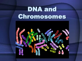
DNA is packaged into chromosomes..
540 likes | 939 Views
DNA is packaged into chromosomes. Unlike DNA chromosomes can be visualized during cell division (light microscope) The word chromosome is derived from Greek chroma (color) and soma (body). Chromosomes are the factors that distinguish one species from another.

DNA is packaged into chromosomes..
E N D
Presentation Transcript
Unlike DNA chromosomes can be visualized during cell division (light microscope) • The word chromosome is derived from Greek chroma (color) and soma (body)
Chromosomes are the factors that distinguish one species from another. • Transmission of genetic info from one generation to the next. • The study of chromosomes and cell division is referred to as cytogenetics • Before 1950 48 chromosomes ??? Sex chromosomes “X” • After 1956 correct chromosomal number 46 Sex chromosomes are “X” and “Y”
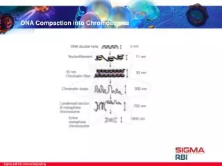
Human Chromosomes Morphology • At the submicroscopic level, chromosomes made up of supercoils of DNA, which has been linked to the tightly coiled network of wiring seen in a solenoid. • Special stains selectively taken up by DNA have enabled each individual chromosome identified.
Each chromosome consist of two identical strands (CHROMATIDS or SISTER CHROMATIDS) • These sister chromatids joined at a primary constriction (CENTROMERE) • Centromeres consist of several hundred kilobases of repetitive DNA and responsible for the movement of chromosomes at cell division
Each centromere divides chromosome into short and long arms; • Short arm : P (petite) • Long arm : Q (Grande) Short arm (p) Long arm (q)
The tip of each chromosome arm is known as the TELOMERE. • Telomeres play a crucial role in sealing the ends of chromosomes and maintaining their structural energy • Telomeres have been highly conserved throughout evolution and in humans they consist of many tandem repeats DNA
Human chromosomesClassification • Morphologically chromosomes are classified according to the position of the centromere • Centrally localized: METACENTRIC • Terminally localized: ACROCENTRIC • Intermediate position: SUBMETACENTRIC
Chromosomes are classified not only to the position of the centromere, but also in their overall length A-G Group A: 1, 2, 3 Group B: 4, 5 Group C: 6, 7, 8, 9, 10, 11, 12, X Group D: 13, 14, 15 Group E: 16, 17, 18 Group F: 19, 20 Group G: 21, 22, Y
Chromosomes as seen at metaphase during cell division TelomereDNA and protein capEnsures replication to tipTether to nuclear membrane Light bandsReplicate early in S phaseLess condensed chromatinTranscriptionally activeGene and GC rich Short armp (petit) CentromereJoins sister chromatids Essential for chromosome segregation at cell division100s of kilobases of repetitive DNA: some non-specific, some chromosome specific Long armq Dark (G) bandsReplicate lateContain condensed chromatinAT rich Telomere
Human chromosome banding patterns seen on light microscopy Chromosome 1 Different chromosome banding resolutions can resolve bands, sub-bands and sub-sub-bands
A pair of homologous chromosomes (number 1) as seen at metaphase Locus (position of a gene or DNA marker) Allele (alternative form of a gene/marker)
Methods of chromosome analysis • Conventional chromosome analysis • High-resolution banding techniques • Molecular chromosome analysis • Florescent in-situ hybridization (FISH) • Comparative genomic hybridization (CGH) • Array based comparative genomic hybridization (array CGH)
Chromosome Preparation • Any tissue with living nucleated cells that undergo division can be used for studying human chromosomes. • Most commonly circulating lymphocytes from peripheral blood are used. • Skin, bone marrow, chorionic villi, cells from amniotic fluid, tumor tissue etc.
Cell culture Peripheral blood sample is added to a small volume of nutrient medium The cells are cultured under sterile conditions at 37C for 3 days, during which they divide Colchicine is added each culture Colchicine has extremely useful property of preventing formation of the spindle, thereby arresting cell division during metaphase Metaphase is the time when the chromosomes are maximally condensed and therefore most visible
Karyotype Analysis • Counting the number of chromosomes • Analysis of the banding pattern of each individual chromosome in selected cells
FISH • This diagnostic tool combines conventional cytogenetics with molecular genetic technology. • The DNA probe s labeled with a fluorochrome which, after hybridization with the patient’s sample allows the region where hybridization occurred to be visualized using fluorescence microscope.
CGH • This technique enabled the detection of regions of allele loss and gene amplification. • Patient DNA (green labeled) and reference DNA (red labeled) samples are mixed and hybridized competitively to normal metaphase chromosomes and an image captured. • If the patient sample contained more DNA from a particular chromosome region than the reference sample that region was identified by an increase in the green to red fluorescence ratio
Clinical cytogenetics is the study of chromosomes, their structure and their inheritance, as applied to the practice of medical genetics. • It has been apparent for nearly 50 years that chromosome abnormalities—microscopically visible changes in the number or structure of chromosomes—could account for a number of clinical conditions that are thus referred to as chromosome disorders.
Chromosome disorders form a major category of genetic disease. They account for a large proportion of all reproductive wastage, congenital malformations, and mental retardation and play an important role in the pathogenesis of malignant disease. • Cytogenetic disorders are present in nearly • 1% of live births, • in about 2% of pregnancies in women older than 35 years who undergo prenatal diagnosis, and • in fully half of all spontaneous first-trimester abortions.
Clinical indications of chromosome analysis • Problems of early growth and development. • Stillbirth and neonatal death. • Fertility problems. • Family history. • Neoplasia • Pregnancy in a woman of advanced age.
Abnormalities of Chromosome Number • A chromosome complement with any chromosome number other than 46 is said to be heteroploid. • An exact multiple of the haploid chromosome number (n) is called euploid, • and any other chromosome number is aneuploid. • In addition to the diploid (2n) number characteristic of normal somatic cells, two other euploid chromosome complements, triploid (3n) and tetraploid (4n), are occasionally observed in clinical material.
Aneuploidy • Aneuploidy is the most common and clinically significant type of human chromosome disorder, occurring in at least 5% of all clinically recognized pregnancies. • Most aneuploid patients have either trisomy or, less often, monosomy • Trisomy can exist for any part of the genome, but trisomy for a whole chromosome is rarely compatible with life. • Monosomy for an entire chromosome is almost always lethal; an important exception is monosomy for the X chromosome, as seen in Turner syndrome.
Although the causes of aneuploidy are not well understood, it is known that the most common chromosomal mechanism is meiotic nondisjunction • Nondisjunction can also occur in a mitotic division after formation of the zygote. If this happens at an early cleavage division, clinically significant mosaicism may result
Abnormalities of Chromosome Structure • Structural rearrangements result from chromosome breakage, followed by reconstitution in an abnormal combination. • overall, structural abnormalities are present in about 1 in 375 newborns. • Structural rearrangements are defined as balanced, if the chromosome set has the normal complement of chromosomal material, • or unbalanced, if there is additional or missing material.
Unbalanced Rearrangements • In unbalanced rearrangements, the phenotype is likely to be abnormal because of deletion, duplication, or both. • Duplication of part of a chromosome leads to partial trisomy; deletion leads to partial monosomy. • Any change that disturbs the normal balance of functional genes can result in abnormal development. • Large deletions or duplications can be detected at the level of routine chromosome banding • Detection of smaller deletions or duplications generally requires more sophisticated analysis, involving FISH or microarray analysis
Deletions • Deletions involve loss of a chromosome segment, resulting in chromosome imbalance • Cytogenetically visible autosomal deletions have an incidence of approximately 1 in 7000 live births. • Smaller, submicroscopic deletions detected by microarray analysis are much more common, but the clinical significance of many such variants has yet to be fully determined !!!
Duplications • Duplications involve gain of a chromosome segment, resulting in chromosome imbalance.
Marker and Ring Chromosomes • Very small, unidentified chromosomes, called marker chromosomes, are occasionally seen in chromosome preparations. • They are usually in addition to the normal chromosome complement and are thus also referred to as supernumerary chromosomes or extra structurally abnormal chromosomes. • Cytogeneticists find it difficult to characterize marker chromosomes specifically by banding, because they are usually so small that the banding pattern is ambiguous or not apparent. • Ring chromosomes are quite rare but have been detected for every human chromosome.
Isochromosomes • An isochromosome is a chromosome in which one arm is missing and the other duplicated in a mirror-image fashion. • The most common isochromosome is an isochromosome of the long arm of the X chromosome, i(Xq), in some individuals with Turner syndrome
Balanced Rearrangements • Chromosomal rearrangements do not usually have a phenotypic effect if they are balanced because all the chromosomal material is present even though it is packaged differently. • It is important to distinguish here between truly balanced rearrangements and those that appear balanced cytogenetically but are really unbalanced at the molecular level. • Even when structural rearrangements are truly balanced, they can pose a threat to the subsequent generation because carriers are likely to produce a high frequency of unbalanced gametes and therefore have an increased risk of having abnormal offspring with unbalanced karyotypes; depending on the specific rearrangement, the risk can range from 1% to as high as 20%.
Inversions • An inversion occurs when a single chromosome undergoes two breaks and is reconstituted with the segment between the breaks inverted. • Inversions are of two types: • paracentric(not including the centromere), in which both breaks occur in one arm; • pericentric (including the centromere), in which there is a break in each arm. • Pericentric inversions are easier to identify cytogenetically because they may change the proportion of the chromosome arms as well as the banding pattern.
Translocations • Translocation involves the exchange of chromosome segments between two, usually nonhomologous, chromosomes. • There are two main types: • reciprocal • Robertsonian.
Reciprocal Translocations • This type of rearrangement results from breakage of nonhomologous chromosomes, with reciprocal exchange of the broken-off segments. • Usually only two chromosomes are involved, and because the exchange is reciprocal, the total chromosome number is unchanged. • Reciprocal translocations are relatively common and are found in approximately 1 in 600 newborns. • Balanced translocations are more commonly found in couples that have had two or more spontaneous abortions and in infertile males than in the general population.
Robertsonian Translocations • This type of rearrangement involves two acrocentric chromosomes that fuse near the centromere region with loss of the short arms • The resulting balanced karyotype has only 45 chromosomes, including the translocation chromosome, which in effect is made up of the long arms of two chromosomes. • Although Robertsonian translocations involving all combinations of the acrocentric chromosomes have been detected, two (13q14q and 14q21q) are relatively common. • Although a carrier of a Robertsonian translocation is phenotypically normal, there is a risk of unbalanced gametes and therefore of unbalanced offspring.